 |


Isthmico-cervical incompetence.
|
|
|
|
Cervical insufficiency has no consistent definition, but is usually characterized by dilatation and shortening of the cervix before the 37th week of gestation in the absence of preterm labor, and is most classically associated with painless, progressive dilatation of the uterine cervix in the second or early third trimester resulting in membrane prolapse, premature rupture of the membranes, mid-trimester pregnancy loss, or preterm birth. Cervical insufficiency arises from the woman’s inability to support a full-term pregnancy due to a functional or structural defect of the cervix. The classic history that raises the suspicion of cervical insufficiency is that of recurrent mid-trimester pregnancy losses. Congenital and acquired cervical abnormalities are causes of cervical insufficiency. Acquired risk factors (cervical trauma) are more common. Cervical trauma may occur during previous childbirth (spontaneous, forceps- or vacuum-assisted delivery), rapid mechanical cervical dilation during gynecologic procedure (eg, uterine scraping), or treatment of cervical intraepithelial neoplasia. Congenital abnormalities include genetic disorders affecting collagen (eg, Ehlers-Danlos syndrome), uterine anomalies, in utero diethylstilbestrol (DES) exposure, and biologic variation.
Depending on the state, several types of cerclage are distinguished.
History-indicated cerclage: insertion of a cerclage as a result of factors in a woman’s obstetric or gynaecological history which increase the risk of spontaneous second-trimester loss or preterm delivery. A history-indicated suture is performed as a prophylactic measure in asymptomatic women and normally inserted electively at 12–14 weeks of gestation.
Ultrasound-indicated cerclage: insertion of a cerclage as a therapeutic measure in cases of cervical length shortening seen on transvaginal ultrasound. Ultrasound-indicated cerclage is performed on asymptomatic women who do not have exposed fetal membranes in the vagina.
Rescue cerclage: insertion of cerclage as a salvage measure in the case of premature cervical dilatation with exposed fetal membranes in the vagina. This may be discovered by ultrasound examination of the cervix or as a result of a speculum/physical examination performed for symptoms such as vaginal discharge, bleeding or ‘sensation of pressure’.
During pregnancy different types of operations may be recommended:
McDonald cerclage, Shirodcar’s suturing of the cervix, Scendy’s operation, Lubimova’s operation and others (Fig. 170, 171, 172).
The McDonald cerclage (Fig. 170) is the most commonly performed and recommended technique. It involves placing a purse-string suture through the cervix as close as possible to the internal os. Care should be taken to avoid the vessels at the 3 and 9 o'clock positions on the cervix. Permanent suture material, such as Mersilene tape or nylon, is used. A second stitch may be placed above the first. The knot usually is tied anteriorly to facilitate removal. The cerclage is removed at the time of labor, in the event of premature rupture of membranes, if infection is suspected, or at 37 weeks.
|
|
|
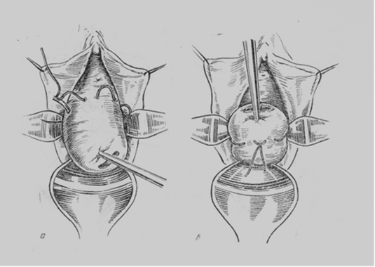
Fig. 170. MacDonald’s operation
2. In the Shirodkar cerclage (Fig. 171), the suture is buried beneath the cervical mucosa, after the bladder is dissected off the anterior cervix. The suture may be left permanently in place, which necessitates cesarean section for delivery, or may be removed to allow for a vaginal delivery. This type of cerclage is associated with more blood loss during placement than the McDonald cerclage and has not been proven to be more effective.
In cases in which a McDonald or Shirodkar cerclage has failed, an abdominal cerclage may be performed before the next pregnancy. Delivery via cesarean section is required after placement of this type of cerclage.

Fig. 171. The diagram of Shirodcar’s operation
3. The Lubimova’s operation (Fig. 172) is the most commonly used in our country. It involves placing of double U-shaped sutures through the cervix in the area of intenal os for constriction of the isthmic site. Lavsan or nylon material is usually used for suturing. The cerclage is removed at the time near the labor, usually at 36-37 weeks of gestation.
After the operation an absolute bed regimen should be prescribed for the patient for a few days, and then she must be under a special care of gynecologist till 36 weeks of gestation. Follow-up of the patient with a cerclage includes frequent cervical examinations either digitally or with ultrasonography. A reduced activity or bed rest as well as abstinence from sexual intercourse may be considered. At 36 week of gestation the sutures are usually taken off and spontaneous delivering will follow.

Fig. 172. The diagram of Lubimova’s operation.
Other option of treatment of isthmico-cervical incompetence is plastic surgery of the cervix (Emmet’s operation) (Fig. 173), which should be done before the next pregnancy.
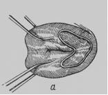
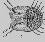
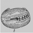
Fig. 173. The diagram of Emmet’s operation
Management of actual preterm labor and delivery (APTL)
WHO definition: Preterm labor is the presence of contractions of sufficient strength and frequency to effect progressive effacement and dilation of the cervix between 22 and 37 weeks' gestation.
Diagnostic criteria to document APTL(20-37w):
• > 4 contractions during 20 minutes
• Cervical dilation > 2 cm in nullipara and > 3 cm in multipara
• Cervical effacement > 80%
The main clinical symptoms are: regular labor pains, cervical dilation for more than 3 cm. Early rupture of membranes, abnormalities of labor pains (weakness, excessive pains, incoordinated pains), intrauterine hypoxia, hemorrhages are characteristic too.
The principles of management of preterm labor are:
· to prevent fetal distress which makes the baby more susceptible to respiratory distress syndrome;
· to prevent birth trauma (Fig. 174 ).
 | |||||||
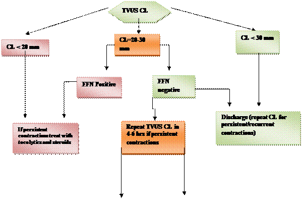 | |||||||
 | |||||||
 | |||||||
Fig. 174 Management of PTL (PTB – preterm birth)
Management of the First stage of Preterm labor: the patient is taken to bed to prevent early rupture of amniotic membranes. A sedative and analgesic therapy should be avoided to prevent depression of the fetal respiratory centre. Therapy to prevent failure of placental microcirculation should be prescribed. Labor should be watched by intensive electronic monitoring. In case of abnormalities of labor pains a special treatment should be administered. If necessary, acceleration of labor by a low rupture of membranes and/or oxytocin infusion should be performed. In case of delay or anticipating a traumatic vaginal delivery, a caesarean section is indicated.
|
|
|
Management of the Second stage of Preterm labor: accurate control of cardiac activity of the fetus, control of uterine contractile activity, prevention of fetal distress. Caesarean section is performed in the presence of obstetric indications. The air passage should be cleared of mucus just after the delivery of head to prevent inspiration of contents and development of asphyxia, atelectasis, pneumonia. The cord is to be clamped immediately at birth to prevent the development of hypervolemia and hyperbilirubinemia.
Premature Infant
A baby born before 37 completed weeks of gestation calculated from the first day of last menstrual period is arbitrarily defined as preterm baby. Babies born before 37 completed weeks usually weigh 2, 500 g and less, the body length is 47 cm and less. The length is more important for diagnosis of prematurity than body weight. It is known that the baby may weigh more than 2, 500 g even when born before 37 completed weeks. On the other hand, baby’s weight may be less than 2500 g even when born after 37 completed weeks of gestation.
Clinical signs of prematurity
The clinical manifestation differs with the degree of prematurity. There are 4 degrees of prematurity:
1st degree — 37-35 weeks of gestation, 2nd — 34-32 weeks, 3rd –31-29 weeks, 4th- less than 29 weeks of gestation. The head and abdomen are relatively large; the skull is soft with wide sutures and posterior fontanelle. The head circumference disproportionately exceeds that of the chest. Normally, the head circumference is larger than the chest circumference at birth and the difference is about 1. 5 cm. The skin is thin, red and shiny, due to lack of subcutaneous fat and covered by plentiful lanugo and vernix caseosa. Muscle tone is poor. The testicles are undescended; the labia minora are exposed because the labia majora are not in contact. There is a tendency of herniation. The nails are not grown right up to the finger tips. Immaturity of organs and systems and physiological mechanisms is a common sign of premature babies. That is why a premature infant is not stable to harmful effects, such as infection, premature anemia, cyanotic attacks. One of the main common and dangerous complication is respiratory distress syndrome (RDS) of a newborn. It is one of the major causes of death in preterm babies born before 34 weeks. RDS almost always occurs in newborns born before 37 wk of gestation: the less term of gestation is, the greater the chance of developing RDS is. The deficiency of lung surfactant, i. e. lecithin and phosphatidyl glycerol, is the principal factor responsible for pulmonary atelectasis leading to hypoxia and acidosis. Rapid, labored, grunting respiration usually develops immediately or within a few hours after delivery, accompanied with suprasternal and substernal retractions and intercostal retractions. The expressiveness of ateletasis and severety of respiratory failure progressively increase. The infant becomes hypoxemic, resulting in metabolic acidosis. If untreated, a severe hypoxemia can result in multiple organ failure and death. However, if the newborn’s ventilation is adequately provided, surfactant poduction will begin and RDS will resolve in 4 or 5 days. Pulmonary surfactant instilled intratracheally reduces the severity of RDS rapidly. Surfactant may be administred immediately after birth to preterm newborns judged to be at a very high risk of developing RDS. Repeated doses of exosurf can be given as needed 12 h apart (up to 3 doses).
|
|
|
Special care is needed for the survival of preterm babies.
The principles that are to be taken for the babies’ special care are:
· to maintain a relatively constant body temperature
· to prevent or to treat atelectasis
· to prevent infection
· to maintain nutrition
· adequate nursing care.
Self Test
1. Termination of pregnancy prior to the 22th week is called:
A. premature labor
B. abortion
C. prematurity
D. postmaturity
2. Profuse bleeding, significant cramping pains in the lower abdomen, expulsion of the fertilizred ovum is:
A. abortion in progress
B. threatened abortion
C. complete abortion
3. Which of the following should be used for treatment of threatened preterm labor?
A. bricanil 0. 5 mg in 400 ml 5% dextrose intravenously
B. 5 units of oxitocin in 400 ml 5% dextrose intravenously
C. vitamin C 300 mg in 400ml 5% dextrose intravenously
4. Premature labor is:
A. termination of pregnancy after 22 weeks but prior to the 37th completed week
B. termination of pregnancy before 38-40 weeks of gestation
C. termination of pregnancy after 14thweek but prior 22 week
5. Which of the following is more important for diagnosis of prematurity?
A. length of the body
B. body weight
6. Which of the following drugs is not indicated for treatment of threatened preterm labor?
A. magnesium sulfate
B. yutopar
C. premarin
D. progesterone IM
7. What is the principal factor responsible for respiratory distress syndrome of a newborn?
A. deficiency in lung surfactant
B. congenital anomaly of the fetus
C. intrauterine infection
8. The sohographic sign of TPTL is cervical length of:
A. > 35mm
B. < 25mm
C. > 45mm
|
|
|
