 |
Calculating the Expected Date of Delivery (E.D.D.) 3 страница
|
|
|
|
Estrogens and calcium are also known to be factors increasing prostaglandin synthesis.
2. Fetal cortisol level. It is possible that fetal cortisol levels and the proper functioning of the fetal adrenal gland induce the spontaneous onset of labor.
3. Progesterone. The increased fetal production of dehydroepiandrosterone (DHEAS) and cortisol may inhibit the conversion of fetal pregnenolone to progesterone, thereby altering the estrogen-progesterone ratio. It is probably the alteration in the estrogen progesterone ratio rather than the fall in the absolute concentration of progesterone, that influences the spontaneous onset of labor.
4. Feto-placental contribution. It has been postulated that due to unknown factors, fetal pituitary gland is stimulated prior to onset of labor, this results in increased release of adrenocorticotropic hormone which stimulates fetal adrenals, then cortisol secretion is increased which results in accelerated production of estrogen and prostaglandin from the placenta. Estrogen is probable to:
• increase the release of oxytocin from maternal pituitary body;
• activate the receptors for oxytocin in the myometrium and deciduas;
• accelerate lysosomal disintegration inside the decidua cells that results in increased prostaglandin synthesis;
• stimulate synthesis of myometrial contraction with protein-actomyosin through activation of adenosine triphosphatase; increase the excitability of the myometrial cell membranes.
5. Oxytocin: It is known that endogenously produced oxytocin plays a role in the spontaneous onset of labor. Oxytocin influences the presence of oxytocin receptors in the myometrium. Myometrial contraction depends on its own readiness to oxytocin action, and this readiness depends on the level of estrogen in blood. There is an increase in oxytocin receptors in the decidua in term of labor. On the other hand, oxytocin stimulates the release of PgF2ά, which occurs in the decidua. The oxytocin level begins to increase just before the labor and reaches the maximum in the second period of labor. Vaginal extension and rupture of the water bag usually result in increasing of endogenous oxytocin level, too.
6. Nervous factors. Labor may also be initiated through nerve parthways. Both alfa- and beta- adrenergic receptors are present in the myometrium: estrogen activates the alfa-receptors and progesterone the beta-receptors to function predominantly. The contractile response is initiated through the alfa-receptors of the postganglionic nerve fibres in and around the cervix and the lower part of the uterus.
Prelabor (syn: premonitory stage). This is a prodromal stage of labor and it may begin two or three weeks before the onset of true labor in primigravidae and a few days before in multiparae. The features are inconsistent and may comprise the following:
• Lightening: a few weeks prior to the onset of labor especially in primigravidae the presenting part sinks into the true pelvis. It may be a gradual process or it may be felt abruptly. This diminishes the fundal height and minimizes the pressure on the diaphragm. The mother experiences a sense of relief from the mechanical cardiorespiratory embarrassment.
|
|
|
• There may be frequent urination or constipation due to mechanical factor — pressure of the presenting part of the fetus.
• Cervical changes: the cervix may become softer, shorter and slightly dilated, especially in primigravidae.
Plentiful mucous discharges from the cervical canal because of cervical changes. These discharges usually appear about twenty-four or forty-eight hours before labor, often mixed with blood. It is the plug, which formerly filled the cervix, closing off the uterine cavity from vagina. The blood comes from the surface left bare by the separation of mucosa. It is called the show.
Appearance of false pain. They usually occur at night, subsiding toward morning.
False pain has the following features:
• dull in nature and usually confined to the lower abdomen and groin;
• weak, irregular and insignificant;
• without any effect on dilatation of the cervix;
• usually relieved by administration of a sedative therapy;
• lessening of body weight, for about 1 kg, due to frеquent urination.
Signs and Symptoms of Normal Labor
• Regular rhythmical uterine contractions, named labor pains. Labor pain contractions occur at decreasing intervals with increasing intensity, causing dilatation of the cervix. These contractions become more powerful and associated with pain. The pains are more often felt in front of the lower abdomen or radiating towards the thighs. The onset may be gradual or abrupt. Initially, contractions last for about 25 seconds, coming as labor pains every 5-7 minutes. Thereafter, the contractions last for 40-55 seconds, coming every 2-3 minutes.
• Effacement and dilatation of the cervix.
• Formation of bag of waters.
Stages of Labor
• The first stage. It starts from the onset of labor pain and ends with full dilatation of the cervix. It is in other words the “cervical stage” of labor (dilation of the cervix). Its average duration is about 12 hours. In multiparae the duration of this stage may be shorter (6-8 hours).
• The second stage. It starts with a full dilatation of the cervix (not from the rupture of the membranes) and ends with expulsion of the fetus from the birth canal. Its average duration is 2 hours in primigravidae and 30 minutes in multiparae. It is in other words the stage of fetus’ expulsion.
• The third stage. It begins after expulsion of the fetus and ends with expulsion of the placenta and membranes (afterbirth). Its average duration is about 30 minutes. The duration, however, is reduced to 5 minutes at active management. It is in other words the stage of afterbirth expulsion.
The First Stage of Labor
Clinical course
One should know that the pacemaker of the uterine contractions is probably situated in the region of the tubal part from where waves of contractions spread downwards. There is a fundal dominance with gradual diminishing contraction wave through midline down to the lower segment, which takes about 10-20 seconds.
The waves of contractions follow a regular pattern. Intraamniotic pressure rises beyond 20 mmHg with the onset of the true labor pains during contraction. Good relaxation occurs in between contractions to bring down the intraamniotic pressure to less than 8 mmHg. It is important to know, that all mentioned features of uterine contractions are very effective only when they are in combination. The first stage of labor is chiefly concerned with the preparation of the birth canal to facilitate expulsion of the fetus in the second stage.
|
|
|
The main events that occur in the 1st stage are:
• regular rhythmical uterine contractions;
• dilatation of the cervix;
• full formation of the lower uterine segment.
Dilatation of the cervix
Prior to the onset of labor, in the premonitory stage there may be a certain amount of dilatation of the cervix, especially in multiparae and in some primigravidae.
Predisposing factors which favour smooth dilatation are:
• cervical ripening;
• fibro-musculo-glandular hypertrophy;
• increased vascularity;
• accumulation of fluid in between collagen fibres.
These are under the action of such hormones as estrogen, progesterone and relaxin.
Three main components constitute a complete cervical examination.
• Cervical ripening is a complex process that ultimately results in physical softening and distensibility of the cervix (see chapter 12). Cervical ripening usually precedes spontaneous labour at term. There is no possibility for effacement and dilatation of immature cervix.
• Effacement is shortening and thinning of the cervix. Effacement of the cervix is a process of thinning which is accomplished during the first stage of labor or even before it. In primiparae, effacement precedes dilatation of the cervix, whereas in multiparae both occur simultaneously. Effacement is expressed as percentage, ranging from 0% (no reduction in length) to 100% (cervix can not be palpable below the fetal presenting part). The cervix is completely effaced (obliterated or unfolded) when it is so changed that only the external os remains. Dilatation now begins or continues.
• Dilation (or dilatation) is a degree of patency of the cervix. Due to regular contractions and retractions of longitudinal and oblique layers of uterine muscles the distraction of circular muscles of the cervix gradually happens. Thus the cervix becomes more and more dilated. In primigravida the cervical canal begins to dilate first in the internal os than in the external one. In multiparae the cervical canal dilates simultaneously in the internal and the external os. Dilatation of the cervix is accompanied by the corresponding stretching of the lower uterine segment. The diameter of the internal os of the cervix is measured in centimeters, from closed to 10 cm, with 10 cm corresponding to complete cervical dilation. After the cervix is completely dilated it rises high and is out of reach.
Actual factors responsible for cervix dilatation are:
• Uterine contraction, retraction and distraction. The longitudinal muscle fibres of the upper segment are attached with circular muscle fibres of the lower segment and upper part of the cervix in a bucket-holding fashion. With each uterine contraction not only the canal is opened up from above down but it also becomes shortened and retracted. There is some coordination between fundal contraction and cervical dilatation called “polarity of uterus”. While the upper segment contracts, retracts and expels the fetus, the lower segment and the cervix dilate in response to the forces of contraction of the upper segment. The eccentric dilation of the cervix is called distraction.
Thus, due to contraction of muscles the fibres become shorter and thicker. Due to retraction the muscles become displaced to each other. And because of both the contraction and retraction, the distraction of the cervical circular muscles takes place (or eccentric tension of the cervical muscles).
• Formation of bag of waters (syn: bag of membranes). The membranes (amnion and chorion) are attached loosely to the deciduas lining the uterine cavity except over the internal os. In vertex presentation, the girdle of contact of the head (that part of the head circumference which first comes in contact with the pelvic brim) being spherical may well fit the wall of the lower uterine segment. Thus, the amniotic cavity is divided into two compartments. The part above the girdle of contact containing the fetus with a bulk of fluid is called hind waters and that below, containing small amount of fluid, is called forewaters. Forewaters are also named a bag of waters, bag of membranes. With the onset of labors forewaters descent into the cervical canal and help to stretch out the cervical canal. (Fig. 121, 122). The best time for the membranes to rupture is when the cervix is completely effaced and dilated, but they may break at any stage, even before the pains begin.
|
|
|
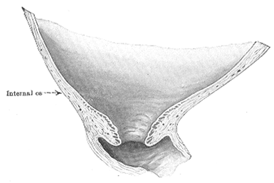
Fig. 121. Effacement and dilatation of the cervix in primipara

Fig. 122. Effacement and dilatation of the cervix in multipara
• Full formation of the lower uterine segment.
• Before the onset of labor there is no complete anatomical or functional division of the uterus. During labor, the demarcation of an active upper segment and a relatively passive lower segment is more pronounced. The wall of the upper segment becomes progressively thickened with progressive thinning of the lower segment. This is pronounced in late first stage, especially after rupture of the membranes and attains its maximum in the second stage. A distinct ridge is produced of the junction of the two, called physiological retraction ring, which should not be confused with the pathological retraction ring — a feature of obstructed labor. The lower segment is thus limited superiorly by the physiological retraction ring and inferiorly by the fibromuscular junction of the cervix and uterus. Anatomically, the lower uterine segment corresponds with the part of the uterus to which the peritoneum is loosely attached to the anterior wall.
During the 1st stage of labor pains become more intensive and longer with the intervals shorter and shorter. The first stage of labor may be divided into 3 phases depending on the character of pain: latent, active and slowing down phases.
Latent phase. During the latent phase, the uterine contractions are typically infrequent, somewhat uncomfortable, and, in some cases, not very strong, but they generate sufficient force to cause slow dilation and some effacement of the cervix. Latent phase of labor begins from the onset of regular labor pain to 4 cm opening of the cervix, the duration of which is about 5 hours in multiparae and 6. 5 hours in primigravidae. The rate of the cervix dilatation is about 0. 35 cm/hour.
Active phase. This phase follows the latent phase and is characterized by rapid cervical dilation, progressive labor pains and descent of the presenting fetal part. The active phase usually begins at about 4-5 cm of cervical dilation and continues until full disclosure. The active phase has a statistical maximum of 11. 7 hours, while the range is from 3-6 hours, dependant of amount of childbirth in previous history. The rate of the cervical dilatation is about 1. 5 — 2 cm/h in multiparae, 1-1. 5 cm/h in nulliparae. Descent of the presenting part begins in the later stage of active dilatation, commencing at 7 to 8 cm in nulliparous and becoming most rapid after 8 cm.
Management of the First Stage of Labor
• Fetal monitoring. Any device should monitor the fetal heart tones immediately after the uterine contraction. During the uterine contraction pulse rate increases by about 10 bmp, and just after contraction it returns to norm. One should know that sudden drop to less than 120 beats per minute (bpm) or increase to above 160 bpm may be an indication of fetal distress. The normal tones are clear and rhythmic, and normalize just after the every uterine contraction.
|
|
|
• Observing the mother’s condition. One should examine mother’s pulse rate, blood pressure, and temperature during the first stage of labor. It should be remembered that during the uterine contraction pulse rate increases by about 10 bmp, and just after contraction it returns to norm. The same refers to blood pressure.
• Monitoring labor pains. Labor pains should be increasing in character during the 1st phase of labor. Estimation of labor pains may depend on uterine palpation together with estimation of tone. The internal hysterography and radiotelemetry may be used as well.
• Observing the cervix dilatation and effacement. It is necessary to make the vaginal examination when the patient is admitted (or with the onset of regular pains), then every 6 hours to understand the changes of the cervix. The rupture of the water membranes, onset of hemorrhages, the descending of the presenting part into the cavity of the pelvis, any complications are indications for the extraordinary internal examination of the patient.
• Pain relief. The main methods of anaesthetizing are:
§ inhalation anesthesia (nitrous oxide);
§ medicamental (no-spa, analgin, baralgin intravenously);
§ epidural anesthesia;
§ electroacupuncture;
§ local anesthesia (pudental blockade).
• Prophylaxis of septic complications (observance of rules of asepsis and antisepsis).
The Second Stage of Labor
Clinical course of the second stage of labor
It is the stage of fetus expulsion. The second stage begins with the complete dilatation of the cervix and ends with the expulsion of the fetus. This stage is concerned with the descent and delivery of the fetus through the birth canal.
With the full dilatation of the cervix, the membranes usually rupture and there is escape of forewaters. After the rupture of bag of waters there is generally a short pause in the pains for the uterus to accommodate itself to the diminished size of its cavity. Then uterine contractions become stronger. The head descends to the pelvic floor. This moment may be diagnosed with the help of Pisckachek method (Fig. 123).
During palpation through the vulva one can reach the fetal head, which means that there is a full dilation of the cervix and head is passed through the pelvic cavity into the pelvic outlet. Delivery of the fetus is accomplished by the downward thrust offered by uterine contractions supplemented by voluntary contraction of the abdominal muscles against the resistance offered by bony and soft tissues of the birth canal. The presenting part of the fetus attains the pelvic floor; the descended head (presenting part of the fetus) presses on the nerves of the rectum and outlet of the pelvis, and the parturient feels like bearing down as at stool. The pains grow stronger and are changed in character, being expulsive. The expulsive force of the uterine contraction is added by voluntary contraction of the abdominal muscles called the “bearing down” efforts.
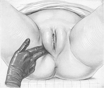
Fig. 123. Determination the rate of advance of the head by pressing in the perineum. The lower pole of the head is palpable through vulva tissues when the head is on the 3rd plane of pelvis.
Thus, it is not only contraction of smooth muscles of the uterus, but also added contraction of skeletal musculature of the abdominal wall. The duration of expulsive pains is about 60-90 seconds with a 1-2 minute interval. Endowed with power of retraction, the fetus is gradually expelled from the uterus against the resistance offered by the pelvic floor. At first the crowning of the presenting part occurs, then disengagement follows. With expulsive pains the perineum bulges more and more, the anus opens wider, the labia separate further and a leading point of the head with neighbouring parts of the scalp become visible. This is called crowning. As the pain disappears the head recedes. When the pelvic floor has relaxed sufficiently, the head rests in the vulva even between pains, and disengagement of the head occurs. Suboccipital fossa states under the lower margin of the pubic arch. Now by supreme efforts and powerful abdominal action, the head is delivered slowly with the base of the occiput rotating around the lower margin of the symphysis pubis — this is extension of the head. So, the extension of the head resulting in delivery of the head is named disengagement. The shoulders appear at the vulva just after external rotation and are delivered spontaneously. The delivery of the fetus trunk is easy.
|
|
|
After the expulsion of the fetus the uterine cavity is permanently reduced in size only to accommodate the afterbirth. On the average, the second stage lasts for approximately 50 minutes in the primigravida and approximately 20 minutes in the multigravida. However, the second stage lasting for 2 hours, especially in the primigravida, is not uncommon. With each contraction, the vulvar opening is dilated by the head. At first the crowning of the presenting part occurs, then disengagement follows. The head then is delivered slowly with the base of the occiput rotating around the lower margin of the symphysis pubis. The shoulders appear at the vulva just after external rotation and are delivered spontaneously. The delivery of the fetus trunk is very easy.
Thus, the main signs of the second stage of labor are:
• Rupture of the bag of waters with escape of liquor amnii;
• Increase of intensity of uterine contractions;
• occurrence of bearing down efforts (syn.: expulsive pains);
• Delivery of the presenting part of the fetus and expulsion of the fetus in whole.
Management of the second stage of labor
The main measures at this stage are observing the rules of asepsis and antisepsis, producing analgesia and anesthesia (discussed previously), preservation of the fetus’ life, protection of perineum, prevention of other obstetric complications (hemorrhages, rupture of uterus, eclampsia, etc. )
Monitoring the fetal heart and medicamental administration for preservation of the fetus’ life are absolutely necessary. One should listen to the heart tones of the child every two or three minutes in the second stage of labor. The electric amplifying stethoscope is also of great use.
Perineum protection. Examination of the pelvic floor after delivery usually shows evidence of injury to all its structures. Rupture of the perineum cannot always be prevented, even with attendance of the best physician and under the most favorable conditions. The pelvic outlet may be weakened by edema, excessive fat deposits, condylomatosis, varicosities, lack of elasticity due to scars or previous operation, age or constitution. A narrow pubic arch, large fetus, mulpresentations, abnormalities of labor pains are the reasons of perineal lacerations. The two principles for protecting the perineum are:
1) slow delivery developing to the ultimate the elasticity of the pelvic floor;
2) delivery of the head in a state of flexion, to present to the parturient passage the smallest circumference of the head.
The physician, having put on a cap, face mask, sterilizes his hands and stands on the right side of the patient, who is lying on her back with the head supported by a thin pillow, knees raised and abducted and with the vulva towards the best light. Every two or three minutes he lifts the sterile towel from the patient’s abdomen, bends his head down and listens to the fetal heart tones. Unless the patient is bearing down too much and the head is advancing too rapidly, he does not interfere by word or act until about 4 cm of the scalp is visible. The rapidity of the descent of the head may be determined easily and safely by pressing the fingers upward and inward along the ramus of the pubis. When the physician decides that the head is coming through too fast, he asks the woman to cease bearing down, to open her mouth and breathe through it until the force of the effort is expended. With each pain the head is allowed to come down to distend the perineum a little more. In gentle fashion the head is permitted to descend until its greatest periphery stands in the distended vulva (the disengagement of the head). It is now delivered in the interval between pains. The head is restrained at the height of the bearing-down effort and the woman is asked to breathe through her mouth, while the physician tucks the clitoris and labia minora behind the occiput under the pubis. After the pain has ceased the woman is asked to bear down and during this effort the distended perineum is slipped back over the baby’s forehead, face and chin. During this maneuver the head is maintained in a state of flexion, not by pressure on the forehead through the perineum but by manipulation of the part that is accessible to the fingers (Fig. 124). During two or three pains a gentle attempt is made to lead the suboccipital part under the pubis by pressing the head back onto the perineum with the finger on the occipital protuberance. When the head has descended far enough to be held, the fingers are applied right at the edge of the frenulum and are used to press the head up under the arch of the pubis. Thus the extension of the head around the point of fixation and delivery of head happen. As soon as the face is delivered the forehead and eyelids are wiped with a cotton sponge, the mucus is squeezed out of the nostrils and wiped out of the mouth. After a minute or two with another pain the patient gently bears down. The external rotation of the head and internal rotation of shoulders occur. Now the child is slightly pressed down as the anterior shoulder comes to the inferior margin of the pubic arch. Then the physician lifts the child up and brings the posterior shoulder over the perineum. Now the rest of the body follows in one long pain without any particular mechanism.
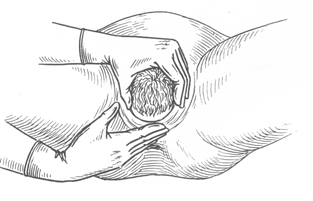
Fig. 124. Protection of the perineum: pressing the fingers upward and toward along the ramus of the pubis.
In case of resistant perineum causing delay in the exit of the head through the vulva, threatened laceration of the perineum, abnormal size of the fetus, obstructed pelvis, delivery of premature baby, the necessity for rapid extraction, perineotomy or episiotomy have to be performed. It is an incision in the perineum that is made either in the midline (perineotomy) (Fig. 125) or starts 2 cm above the midline but is directed laterally away from the rectum (a mediolateral episiotomy) (Fig. 126)
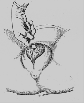
Fig. 125. Direct perineotomy
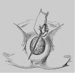
Fig. 126. Episiotomy
The Third Stage of Labor
It is the shortest stage of labor. It starts from the expulsion of the fetus and ends with expulsion of the placenta and membranes. The expelled placenta with membranes is called afterbirth.
The duration of this phase is not more than 30 minutes. But more often expulsion of afterbirth takes place during 5–10 minutes after fetus expulsion.
The patient has uterine contractions in this phase, which lead to separation of the placenta, descending and expulsion with membranes.
The mechanism of placental separation. Marked retraction reduces effectively the surface at the placental site to about its half. However, as the placenta is inelastic, it cannot keep pace with such diminution of the uterus after the fetus delivery. A sharing force is instituted between the placenta and the placental site, which brings about its ultimate separation. The plane of separation runs through deep spongy layer of decidua basalis so that a variable thickness of decidua covers the maternal surface of the separated placenta. There are two ways of separation of the placenta.
Central separation (by Schults). Detachment of the placenta from its uterine attachment starts in the centre resulting in opening up of few uterine sinuses and accumulation of blood behind the placenta (retro placental hematoma). With increasing contraction, more and more detachment occurs facilitated by weight of the placenta and retro placental blood until the whole of the placenta gets detached. There is no any hemorrhage until delivering of the whole placenta.
Marginal separation (by Duncan). Separation starts at the margin, as it is mostly unsupported. With progressive uterine contraction, more and more areas of the placenta get separated. There is some bleeding during the separation of the placenta.
One should know the volume of physiological hemorrhage, which takes place in the normal third period. It is about 0. 5% of body weight, but not more than 400 ml. For example, the patient’s body weight is 70 kg, so physiological hemorrhage in the third period of labor is about 350 ml. It is the volume of blood, which was the content of the intrafibre space of the placenta.
After placental separation innumerable torn sinuses, which have free circulation of blood from the uterine and ovarian vessels, have to be obliterated. The occlusion is effected by complete retraction whereby the arterioles, as they pass through the interlacing intermediate layer of the myometrium, are literally clamped.
The second mechanism to prevent bleeding is thrombosis which occurs to occlude the torn sinuses, a phenomenon which is facilitated by hypercoagulable state of pregnancy. Apposition of the walls of the uterus following expulsion of the placenta (myotamponade) also contributes to minimization of blood loss.
Management of the Third Stage of Labor
This phase of labor is very important due to the possibility of hemorrhage and septic complication after labor. Catheterization of the bladder just after the fetus expulsion can facilitate the separation of the afterbirth and present bleedings.
The main problem of this phase is to prevent bleeding. One should observe the following signs of placenta separation.
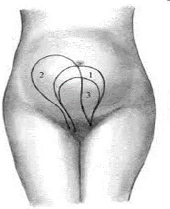
1. Shredder sign. The fundus of the uterus rises above the umbilicus, the uterus shape becomes stretched, the fundus deviates to the right side (Fig. 127): change in shape of the uterus from flat (discoid) to round (globular), the fundus of the uterus rises above the umbilicus, the uterus shape becomes stretched, the fundus deviates to the right side.
|
|
|


