 |


Criteria for Discharging Mothers from the Maternity Home.
|
|
|
|
• A satisfactory state of patient’s health;
• A normal body temperature for the last 3 days;
• Normal characteristics of pulse (rate, rhythm, tension, volume);
• Uniformly soft mammary glands (hypogalactia and agalactia are not the indication for delay of patient in the clinic);
• Cicatrization of wounds of soft maternal passages;
• Moulded cervix of uterus, allowing not more than 1 finger in the area of the internal os;
• Body of uterus is not more than 10-12 cm in length, painless, mobile;
• Absence of induration and morbidity in uterine appendages and parametrium;
• Lochia serosa, without smell;
• Absence of pathological signs in blood test.
Soon after leaving the maternity home, the child should be followed-up in the pediatric clinic. General hygienic measures of the mother should include regular cleaning of the body surface (taking shower) until complete epithelization of wounds on the surface of uterus (at the end of 3rd week); afterwards bath is allowed.
The sexual life is allowed not earlier than on the 6-8th week of afterbirth period, when complete involution of internal genital organs occurs. It is necessary to note that restoration of menstrual function and occurrence of the next pregnancy in puerperal period have individual features. In feeding mothers lactation amenorrhea proceeds during 4-7 months and in some women during the whole period of breast-feeding. It is necessary to know that the minority of breastfeeding women begins to menstruate by the end of puerperal period. With occurrence of menstrual function the possibility of pregnancy onset appears. And as ovulation precedes the menstruation, pregnancy can come without menstruation. Puerperal patients during their stay in the maternity home must get recommendations in regard to modern means of contraception; booklets on the given topic should be handed to them.
At significant flaccidity of the abdominal wall, bearing of abdominal bandage should be recommended, as well as continuation of therapeutic exercise (its complex should be mastered in the maternity home). The neglect of these recommendations results in formation of pendulous abdomen and visceroptosis.
Self Test
1. The average blood loss at vaginal delivery is:
A. 100 ml
B. 200 ml
C. 300 ml
D. 0. 5 % of body weight
2. Blood transfusion therapy in the early puerperal period should be started depending on:
A. amount of blood loss
B. systolic blood pressure
C. diastolic blood pressure
D. pulse pressure
E. pulse rate
3. Colostrum differs from mature milk in:
A. less proteins
B. more immunoglobulins
C. more fat
D. more lactose
E. fewer cells
4. Which of the following is contraindicated to breast-feeding women?
A. ampicillin
B. lithium
C. metronidazole
D. propylthiouracil
5. By the end of the 5th day of puerperal period the uterus is:
A. on the midline between the symphysis pubis and umbilicus
B on the level of the umbilicus
|
|
|
C. 2 cm above the umbilicus
D. 2 cm below the umbilicus
6. Identify the number of independent secretory glands in the lactating breast:
A. 1
B. 3-4
C. 5-7
D. 8-10
E. > 15
7. Which of the following is of importance in galactopoesis?
A. oxytocin and prolactin
B. oxytocin and estrogen
C. estrogen and progesterone
D. estrogen and cortisol
8. The duration of early puerperal period is:
A. 2 hours
B. 30 minutes
C. 2 days
D. 6-8 hours
9. The duration of the late puerperal period is:
A. 6–8 weeks
B. 2 days
C. 30 days
D. 2 months
10. The length of the uterus diminishes daily by:
A. 1-1. 5 cm
B. 3-4 cm
C. 0. 5 cm
CHAPTER 17. The course of pregnancy and labor in breech presentations
Presentation when the pelvic extremity of the fetus lies at the brim and the cephalic extremity at uterus fundus is called breech. The birth in pelvic presentation is recognized as pathological because of the increased risk to the child during delivery, mainly due to the compression of the umbilical cord at the time of the birth of the head. The compression of the umbilical cord can also occur at the birth of the shoulders of the fetus.
Classification of breech presentations
There are two types of breech presentations depending on the attitude: flexed (breech) and deflexed (footling) presentations.
Breech presentations may be complete and incomplete. At complete breech presentation the attitude of full flexion is maintained. Thighs are flexed at the hips and legs — at the knees. The presenting part consists of two buttocks, external genitalia and two feet. (Fig. 134). It is commonly present in multiparae.

Fig. 134. Complete breech presentation
At incomplete presentation the attitude of flexion is not maintained. It is named a frank breech ptesentation. In this condition the thighs are flexed on the trunk and the knee joints are unbended. The presenting part consists of two buttocks and external genitalia only. (Fig. 135).

Fig. 135. Incomplete (frank) breech presentation
It is commonly present in primigravidae (about 70%). The increased prevalence in primigravidae is due to a tight abdominal wall, good uterine tone.
Footling presentation may be complete, incomplete and knee.
Complete footling presentation: both the thighs and the legs are extended bringing the legs to present at the brim (Fig. 136).

Fig. 136. Complete footling presentation
Incomplete footling presentation: the presenting part is one of the legs, while another is flexed and located superiorly.
Knee presentation: thighs are extended but the knees are flexed, bringing the knees down to present at the brim. These varieties are not common.
Incidence. The incidence is about 5% in 34 weeks and 3% at term. Thus in 3 cases out of 4 the spontaneous correction into vertex presentation occurs by the period of 34 weeks.
Factors predisposing to the formation of pelvic presentation:
• maternal: congenital abnormalities of the uterus, tumors of the uterus body, a scar on the uterus, contracted pelvis, a decrease or increase uterine tonicity, numerous pregnancies in previous history and others;
• fetal: congenital anomalies of fetal development, prematurity, multiple gestation;
• placental: placenta previa, localization of placenta at the uterus fundus (too high), polyhydramnios, short umbilical cord.
|
|
|
There is a high incidence of breech presentation in early weeks of pregnancy. Presentation of the fetus becomes stabilized to 36 weeks.
The Diagnosis of Breech Presentations
It is based on palpation (external and internal), auscultation of the fetus heart sounds, ultrasound examination.
Fundal grip: one can find large, globular, hard part of the fetus with clear contours in the fundal area. If it is ballotable, it confirms that it is the head.
Third grip: one can find a broad, soft and irregular mass suggestive of breech presenting the pelvis brim.
Auscultation of the fetus heart sounds: they are better heard above the umbilicus, on the right or left side depending on position.
Bimanual examination: one can find irregular presenting part without any fontanelles, sutures. After the rupture of the water membrane one can find buttocks, two ischial tuberosities, the coccyx, the rough posterior surface of the sacrum, the anus, and one or both feet.
The anus is distinguished from the mouth by the presence of the elastic sphincter, the absence of the ridges of the gums, and the possible appearance of meconium on the examining finger. The groin differs from the axilla in the absence of ribs. The foot is distinguished from the hand by all the toes being of nearly the same length, absence of thumb, and presence of the heel.
The Course of Pregnancy in Breech Presentations and Management of pregnant women with breech presentations
Pregnancy may be complicated by premature rupture of water membranes, premature labor.
It is possible to change the fetus presentation with the help of external rotation of the fetus. The external cephalic version may be performed closely to the 35-37th week of pregnancy.
Contraindications for external cephalic version:
• fetal malformations,
• threatened preterm labor,
• infertility and miscarriage in previous history,
• Hypertension of pregnancy, preeclampsia-eclampsia syndrome,
• placenta previa,
• abnormalities of the uterus,
• oligohydramnios or polyhydramnios,
• multiple fetuses,
• narrow pelvis,
• severe extragenital diseases,
• low location of the fetus,
• obesity,
• localization of the placenta on the anterior wall of the uterus.
The techniques of external version: just before the procedure, the US should be made to determine the position of the baby, the location of the placenta and the amount of amniotic fluid in the uterus. After the ultrasound assessment and fetal testing, informed consent should be obtained, taking in to account the information gathered from the fetal testing and ultrasound. The patient lies on her back, her legs are slightly divorced in the hip joints and bent at the knee. Practitioner place his hands (one hand is placed on the fetal head and the other is on the fetal buttocks) on the maternal abdomen to gently turn the fetus from breech to cephalic presentation. Soft pushing on the abdomen can lead the child to the " upside down" position. The external version has a high success rate.
Possible complications of external version are:
• placental abruption,
• premature discharge of forewaters,
• rupture of the uterus,
• amniotic fluid embolism,
• premature labor,
• intrauterine fetal distress and death of the fetus.
When carrying out an external cephalic version, an informed consent of the patient is mandatory.
On the other hand special gymnastics may be administered to the patients with breech presentation with the aim of spontaneous turning of the fetus.
Contraindications for gymnastic exercises are similar to those for an externalcephalic version.
At 36-38 weeks with breech presentation ultrasound is used to determine the degree of extension of the fetal head. The angle of extension of the head is determined between the occipital bone and the vertebral column.
|
|
|
In this case, there are 4 variants of the position of the head in the breech presentation:
• the head is bent, the angle is more than 110 degrees;
• the head is slightly unbent, the angle is from 100 to 110 degrees – the 1st degree of extension;
• head moderately unbent, angle from 90 to 100 degrees – the 2nd degree of extension;
•excessive extension of the head, angle less than 90 degrees - the 3rd degree of extension.
At 38-39 weeks patient should be hospitalised to to choose the mode of delivery and to prepare for labor/delivery. In the hospital the following examination should be performed:
• common laboratory tests,
• determination cervical ripening by Bishop scale,
• US: to evaluate expectant body weight of the fetus, location of the placenta, lie and position of the fetus, degree of extension of the head, presence of umbilical cord pathology, etc;
• non-stress test (CTG).
Indications for cesarean section in the planned order with full term pregnancy:
• primigravida older than 30 years,
• obesity,
• severe preeclampsia,
• extragenital diseases requiring substitution of expulsive pains,
• contracted pelvis of any degree,
• the estimated weight of the fetus is more than 3, 600 grams
• fetal hypotrophy of II-III degree,
• fetus distress (diagnosed by CTG, Doppler etc),
• extension of the head of grade 2 - 3 by ultrasound,
• post term pregnancy,
• footling presentation,
• Rhesus incompatibility,
• multiple pregnancy with breech presentation
• pregnancy after artificial reproductive techniques,
• scar on the uterus after previous cesarean section, myomectomy,
• complicated anamnesis: stillbirths, habitual miscarriage,
• refusal of a woman from natural childbirth.
Indications for delivery through maternal passages:
• Good condition of mother
• Good fetus condition
• Mature (ready for labor) cervix
• The maternal pelvis sizes and the fetus sizes correspond to each other.
In most countries the basic method of delivery in breech presentation is a cesarean section in a planned manner.
Mechanism of Labor in Breech Presentation
• The 1st moment is the engagement of the hips. The hips of the fetus enter the pelvis so that the bitrochanteric diameter aligns with one of the oblique diameters of the mother’s pelvis. The hips of the fetus descend into the mother’s pelvis with the leading buttock below the aftercoming buttock. The anterior buttock is thus the leading point and a swelling is formed on it (Fig. 137).

Fig. 137. Breech presentation. The 1st moment is the engagement of the hips.
• The 2nd moment is internal rotation of the buttocks. The buttocks are rotated in the pelvic cavity, the bitrochanteric diameter aligns with the anteroposterior diameter of the pelvis so that the anterior buttock rests (takes up position) against the pubis and the posterior one faces the sacrum.
• The 3rd moment is crowning the buttocks. At first, the anterior iliac bone of the fetus is fixed to the inferior margin of the pubic arch, which works as a fulcrum, around which the fetal body is flexed to a high degree, and the posterior buttock is delivered. Then the anterior buttock rests too. The buttocks are delivered together with the legs in complete breech presentation, and the legs prolapse after the delivery of the body in frank breech presentation. After the lower end of the body has been delivered, it is deflexed and by a few contractions is delivered to the navel and then to the lower angle of the shoulder blades. The body is slightly turned with its back anteriorly. (Fig. 138).
|
|
|
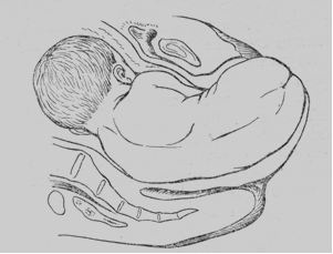
Fig. 138. Breech presentation. The 3rd moment is crowning of the buttocks
• The 4th moment is shoulders delivery. The transverse diameter of the shoulders aligns with the oblique diameter of the mother’s pelvis (the same as with the passage of the buttocks), but later turns to align with the anteroposterior diameter of the pelvis. The anterior shoulder passes down under the pubic arch, and is fixed to the inferior margin of the arch, while the posterior shoulder sweeps over the perineum (Fig. 139).

Fig. 139. Breech presentation. The 4th moment is shoulders delivery.
5. The 5th moment is head delivery. The head enters the pelvis in transverse diameter of the pelvis, or in one of oblique diameters. The engaging diameter of the head is small oblique. As it descends, it rotates into the antero-posterior diameter of the outlet, the occiput passing to the front and coming under the pubic arch, while the face passes over the surface of the sacrum. The suboccipital fossa is fixed to the arch, and rotation around the point of fixation occurs. The chin, face, brow and vertex then appear in that order over the perineum. (Fig. 140).
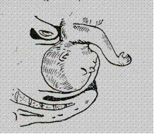
Fig. 140. Breech presentation. The 5th moment is head delivery
Typical Complications in labor with Breech Presentations
It is necessary to understand that due to pressing of the umbilical cord with descending head asphyxia may develop. So, if the head passes the pelvis for about 4-4. 5 minutes, it is not dangerous, but if it lasts more than 5 minutes, it may lead to intrauterine death of the fetus. Concerning the birth of the aftercoming head, it is of importance that the head should be kept in a flexed condition all the time. Extension of the head means its delivery with large diameter that is why its further descending is delayed. It is this delay that constitutes the main risk in breech presentation. The usual fetal mortality both in primigravidae and multiparous women is about 8-10%. The principal risks to the child are intracranial haemorrhage and asphyxia due to the head extension and delay in the pelvic cavity. In addition to these risks, the fetus may receive injuries to limbs, joints and nerve trunks during the efforts to deliver the head or breech, e. g. fracture or dislocation, hematoma of sternocleidomastoid muscle.
Besides, a hand (or both hands) may be thrown back which leads to delay of shoulder and head delivery. Three degrees of throwing a hand (or hands) back may be distinguished:
The 1st degree — a hand (or hands) to the face;
The 2nd degree — a hand (or hands) to the ear;
The 3rd degree — a hand (hands) to the occiput (Fig. 141)
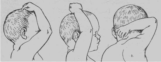
Fig. 141. Breech presentation. Three degrees of sinking back of the hand (or hands)
Manual Assistance in Breesh Presenations
Classical Manual Assistance in Breech Presentations (with Extended Hand and Head)
In case of throwing a hand (or hands) back a special maneuver called classical manual removing of extended hand (or hands) must be used to help the delivery. At first the posterior hand has to be delivered. The obstetrician’s fingers (of the right hand if the infant’s back is directed towards the left side of the pelvis, and of the left hand if the back is directed towards the opposite side) are inserted into the posterior pelvis until they reach the arm and the forearm of the fetus. To help this, the obstetrician’s another hand tries to lift the fetus’ legs upwards. (Fig. 142).

Fig. 142. Classical manual assistance in breech presentations. The operator’s fingers are inserted into the posterior pelvis until they reach the arm and the forearm of the fetus.
Thus, the arm and forearm are swept across the ventral surface of the infant and out of the vagina. So, the fetus’ posterior hand is delivered. If the fetus anterior hand is thrown back too, the obstetrician should rotate the infant body under the arch by 180 degrees to make the anterior arm posterior (Fig. 143).

Fig 143. Classical manual assistance in breech presentations. Rotation of the infant body under the pubic arch.
And then the whole process must be repeated. More often babies are lost during extraction of the hand than at any other stage of breech delivery. Anoxia develops rapidly because the cord is compressed between the head and the pelvic brim and the infant cannot breathe yet. Unless the mouth is free within three or five minutes, anoxic brain damage may occur. Because of anoxia danger, delivery of the head should be done quickly and carefully, with the least forceful and frantic attempts, which may be more damaging than helpful. It is here that understanding of mechanism of breech delivery, experience, and abilities are of greatest importance, because there is no opportunity to seek consultation, and the obstetrician must know exactly how to preceed.
|
|
|
In most instances the head descends through the inlet and rotates to an anteroposterior position and can be delivered without difficulty, but occasionally it delays and cannot enter the pelvic cavity because of extention.
If the fetus head is extended the Moriso-Levre-Le-Shapell maneuver must be used. The obstetrician’s forefinger is inserted into the vagina, and then it should be inserted into the fetus mouth, and pressed against the mandible until the head is flexed. At this time the delivered fetus body is situated on the operator’s forearm like on horseback. Thus, the head is flexed. (Fig. 144). Then the external operator’s hand is located on the fetus shoulders with the 2nd finger on one shoulder and 3rd — on another. Then the head is delivered with the help of traction. The force of traction is at first downward until the mouth appears at the outlet, and then the fetus body is raised while the head revolves around the pubic arch and is delivered. After the forehead crosses the perineum, the rest of the head can usually be delivered without difficulty. The maneuver which helps to deliver the sinking back hand (or hands) and extended head of the fetus is called a classic manual maneuver in breech deliveries.
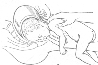
Fig. 144. Classical manual assistance in breech presentations. The Moriso-Levre-La-Shapel maneuver.
Manual assistance in frank breech presentations (Tsovyanov’s method №1)
The method of conducting labor in frank (breech) presentation suggested by N. A. Tsovyanov is now widely used in obstetric practice in Ukraine to reduce the stillbirth rate. The method is based on preservation of normal relation between the parts of the fetal body to prevent possible complications such as extension of the arms or extension of the head. In normal frank breech presentation, the legs are extended along the body and press the flexed arms to the fetal chest. The feet of the extended legs reach the face and thus promote the flexion of the head with the chin resting against the chest.
According to the proposed method, the normal arrangement of the fetal parts is preserved by the extended legs which are pressed against the body and are not allowed to be delivered prematurely.
The normal arrangement of the body parts is also important for the dilation of the birth canal to the extent required to pass the aftercoming head. The girth of the body with the arms flexed on the chest and the extended legs (at the shoulder level) is considerably greater then the girth of the head (42 cm against 32-34 cm), and the latter is therefore delivered without any obstacle.
Manual assistance according to Tsovyanov in frank breech presentation consists in the following. When the hips are delivered, they are grasped by hands as follows: the thumbs are pressed against the legs flexed on the abdomen, while the other fingers of both hands hold the fetus by the sacrum. This position of the fingers prevents: 1) premature prolapse of the legs; 2) hanging down of the delivered body. The delivered body is lifted in the direction of the pelvic axis. The fetus body being expelled from the uterus, the obstetrician moves his hands in the direction of the pudendal cleft of the parturient, pressing carefully the flexed legs against the abdomen by the thumbs; the other fingers slide higher along fetal back (Fig. 145). It is necessary that the legs should not prolapse before the shoulder girdle has been delivered. As soon as the shoulders are delivered, the arms usually prolapse spontaneously. If it fails, they are released as follows. Without changing the position of the obstetrician hands, the shoulders are turned to align with the anteroposterior diameter of the mother’s pelvis and the body is flexed posteriorly: the anterior arm is delivered from under the pubic arch. The body is then lifted anteriorly and the posterior arm is delivered over the perineum.
The heels of the fetus prolapse simultaneously with the posterior arm, and the chin and the mouth show at the pudendal cleft. The head is delivered spontaneously by strong expulsive efforts, while the body should at this moment be lifted anteriorly. If the delivery of the head is delayed, it should be assisted by the the Moriso-Levre-Le-Shapell maneuver (the obstetrician’s hand is inserted into the vagina, it should be inserted into the fetus’ mouth and press the mandible while the head is flexed. The external obstetrician’s hand is situated on the fetus’ shoulders with the 2nd finger on one shoulder and the 3rd — on the other. Then the head is delivered with the help of traction).
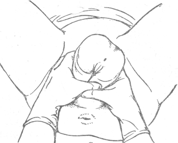
Fig. 145. Tsovyanov’s method №1. Grasping of the hips.
Manual Assistance in Footling Presentations (Tsovyanov’s method № 2)
The incidence of complicated labor in footling presentations is higher than in frank or complete breech presentation.
Complications occur because the forthcoming legs cannot dilate the birth canal to the extent required to pass the bulky shoulders and the head. The head is therefore often deflexed and the arms extended in footling presentations. Not infrequently the head is compressed by the contractions of the uterine cervix.
These complications can be precluded if, by the moment of expulsion of the shoulders, the uterine os is opened completely.
To ensure this, Tsovyanov suggested that a method should be used by which the legs are retained in the vagina until the uterine os has completely opened.
If the diagnosis of footling presentation has been confirmed by the vaginal examination, the obstetrician covers the external genitalia with a sterile napkin, places the palm of the hand over it and prevents premature expulsion of the legs. This ensures full opening of the uterine os because the fetus “squats” to assume the complete breech presentation. As the fetus further moves along the birth canal, it presses the sacral nerve plexus to intensify the uterine and abdominal muscle contractions.
Complete opening of the cervical os is evidenced by protrusion of the perineal area under the thrust of the fetal hips, by the opening anus, frequent and strong expulsive efforts, and the presence of the contraction ring at the level of 10-12 cm above the symphysis. Under the downward thrust of the descending hips the pudendal cleft opens and the legs overcome the resistance of the obstetrician hand to show from under it. If the signs of full dilation are obvious, the obstetrician removes his hand and further labor is conducted as described for breech birth.
|
|
|
