 |


Management in labor. Intrauterine growth retardation (syn: dysmaturity, small-for-gestational-age-infant, iugr). Management of IUGR
|
|
|
|
Management in labor
· Avoid supine position of mother
· To arrest augmentation of labor pains with Oxitocin
· In case of intrapartum fetal distress:
· To make VE and evaluate obstetrical situation:
§ In the 1st stage of labor cesarean section is indicated
§ In the 2nd stage – forceps delivery/ vacuum delivery/ extraction of the fetus by podalic end (in breech presentation).
Intrauterine growth retardation (syn: dysmaturity, small-for-gestational-age-infant, iugr)
Any infant whose weight is below the 10th percentile for gestational age is named a small-for-gestational age infant.
Etiology
An infant may be small at birth because of genetic factors. Nongenetic factors that can retard intrauterine growth are usually not apparent before 32 to 34 wk gestation and include placental insufficiency that often results from maternal disease involving the small blood vessels (as in preeclampsia, primary hypertension, renal disease, longstanding diabetes); placental involution accompanying postmaturity; infectious agents, such as cytomegalovirus, rubella virus, virus of toxoplasmosis.
An infant may also be small for gestational age if the mother is a narcotic addict, or a heavy user of alcohol, or to a lesser degree, if she smoked cigarettes during pregnancy.
The syndrome of intrauterine growth retardation (IGR) can be encountered in two forms:
· symmetric - (incidence is 10-30%) - develops from the early terms of pregnancy and is characterized by proportional lag of body mass and length of fetus. Reasons are various: intrauterine infections, chromosomal anomalies, inadequate nutrition of mother, genetic syndromes, mother’s harmful habits, radiation, medicinal therapy, hypoxic diseases in mother (heart diseases of dark blue type, severe asthma);
· asymmetric - (incidence is 70-90%) is characterized by lag of body mass at normal length of fetus (hypotrophy) with delay in development of some organs (liver, other parenchymatous organs). It is more common in the III trimester of pregnancy on a background of pregnancy complications (EPH-complex, multiple pregnancy, defects of development of placenta, bleeding in the III trimester of regnancy) and extragenital diseases.
Degrees of IGR at Pregnancy
I degree - lag in development of fetus up to 2 weeks.
II degree - lag in development of fetus up to 2-4 weeks.
III degree - lag in development of fetus over 4 weeks (irreversible changes and intrauterine fetal death are possible).
At IGR the delay or absence of uterus growth (disparity to gestational age of fetus) and absence of increase of body mass of pregnant are originally marked, then symptoms of chronic intrauterine fetal hypoxia occur. The delay of development of fetus may be accompanied by symptoms of threatened interruption of pregnancy. The first signs of IGR can appear in 18-19 or 24-26 wk. By 28-29 wk of pregnancy the delay of fetus development, as a rule, is symmetric, i. e. deficit of body mass and length of the fetus is available. This type of intrauterine growth retardation is less favourable.
|
|
|
The onset of syndrome in 32 weeks of pregnancy is more typical of asymmetric form.
If there is only a mass deficit at full-term newborn, it means that a factor causing retardation of fetal growth affected during the last 2-3 months of pregnancy.
After delivery of fetus the type of intrauterine growth retardation of the fetus is determined by a clinical picture for making a prognosis of the course of early neonatal period.
A Hypotrophic Type, or Asymmetric Form of IGR
At 1st degree thinning of subcutaneous fat, decline of turgor of tissues occur.
At 2nd degree the skin of a child is dry, pale, with peeling and chaps, the subcutaneous fat is thin. The course of neonatal period is usually complicated by asphyxia, metabolic disturbances. Early neurological disorders such as syndrome of hyperexcitability or suppression of the central nervous system come to light.
At 3rd degree of asymmetric form of IGR the skin is wrinkled, folded, dry, pale, with chaps. The subcutaneous fat is absent, muscular mass is diminished. In early neonatal period there is usually a hemorrhagic syndrome, anemias, neurological, cardiac, infectious complications.
A Hypoplastic Type, or Symmetric Form of IGR
Children at this variant of IGR look proportionally built but little. The degree of IGR is determined by deficit of body length and circumference of head in relation to term of pregnancy. In early neonatal period hypoglycemia, hyperbilirubinemia, infectious complications are common.
A Dysontogenetic Type
This type is encountered at combination of delay of IGR with vices of development. The state of children at this variant is determined by presence of hereditary diseases and vices of development.
Management of IUGR
Despite numerous approaches to managing IGR, there are no effective treatments to improve the growth pattern of a fetus. The universally available therapeutic option that shows improvement in outcome includes the antenatal administration of steroids in preterm pregnancies and delivery at an institution with a neonatal care unit that is able to deal with the management complexities of the growth‐ restricted neonate. Antenatal steroids should be given to any growth‐ restricted fetus whose delivery is expected before 34 weeks' gestation. Betamethasone (given as a combination of betamethasone sodium phosphate and betamethasone acetate), is administered as two doses of 12mg given intramuscularly, 24 hours apart. Dexamethasone sodium phosphate is administered as four doses of 6mg given intramuscularly, 12 hours apart. As there are no effective treatments to reverse fetal growth restriction, prenatal management is aimed primarily at determining the ideal timing and mode of delivery. This assessment must be individualised, depending on several variables: gestational age of the fetus, maternal health, severity of growth restriction and fetal well‐ being. At term < 34 weeks conduct weekly biophysical profile and umbilical artery Doppler. If growth plateaus or stops, decision would depend on Doppler investigation results: if abnormal uterine artery, MCA (middle cerebral artery), and DV (ductus venosus) Doppler studies and abnormal NST: urgent delivery is more preferable. Cesarean section for fetus with IGR is associated with a lower rate of respiratory distress syndrome, neonatal seizures, and death.
|
|
|
Prognosis
Perinatal asphyxia is the most serious potential complication for infants with IGR because of placental insufficiency, and if it can be avoided, a neurologic prognosis is quite favourable for these infants. Infants with IGR due to genetic factors, congenital infections, or maternal drug abuse often have a worse prognosis, which depends on specific diagnosis.
Self Test
1. Which of the following types of fetal hypoxia is not distinguished?
A. tissue hypoxia
B. anemic hypoxia
C. hypoxic hypoxia
D. circulatory hypoxia
E. respiratory hypoxia
2. Which is the earliest symptom of intrauterine hypoxia of the fetus?
A. tachycardia
B. bradycardia
C. decreased fetal mobility
3. Summary score in biophysical profile from 8 to 10 is
A. a normal fetal condition.
B. a sign of fetal hypoxia.
4. Summary score in biophysical profile from 4 to 6 is
A. a normal fetal condition.
B. a sign of fetal hypoxia.
5. Which of the following signs should be used for evaluation of the status of neonate with asphyxia?
A. natural respiration, heart rate, colour of the skin
B. bradycardia, weak reflexes and satisfactory muscular tone
C. achilles reflex, colour of the skin, heart rate
CHAPTER 36. INJURIES TO A NEWBORN
Birth injuries remain an important cause of perinatal morbidity and mortality. Birth injuries are those which occur during birth of a child.
Incidence.
The incidence of any injuries of newborn is about 2%.
Classification.
Birth injuries may be classified according to localization and etiology.
1. Injuries to the head:
- cephalohematoma
- scalp injuries
- skull fracture
2. Intracranial hemorrhages:
· Traumatic or anoxic:
- extradural hemorrhage
- subdural hemorrhage
- intraventricular hemorrhage
- intracerebral hemorrhage
3. Injuries to skin and subcutaneous tissues
4. Muscles injuries
5. Nerve injuries
6. Injuries to bones (fractures and dislocations)
7. Visceral injuries
Injuries to the Head
Cephalohematoma
It is an effusion of blood in between the pericranium and a flat bone of the skull, usually unilateral and over the parietal bone. It is due to rupture of a small emissary vein from the skull and may be associated with fracture of the skull bone. This may be caused by forceps delivery but may also be met with following a normal labor. The swelling is limited by the suture lines of the skull as the pericranium is fixed to the margins of the bone. It is circumscribed, soft, fluctuant and incompressible. There may be underlying fracture of the skull. In the course of time, a hard sharp edge can be felt surrounding the swelling due to organization of the blood. The condition may be confused with caput succedaneum or meningocele. Meningocele always lies over a suture line or fontanelle and it bulges at crying. Prognosis is good. The blood is absorbed in the course of time (6-8 weeks) leaving an entirely normal skull. Rarely suppuration occurs. No active treatment is necessary. Prevention of infection and avoidance of trauma are important.
Scalp Injuries
Minor injuries of the scalp such as abrasion in forceps delivery (tip of the blades), incised wound inflicted during cesarean section or episiotomy may be met with. On occasion, the incised wound may cause brisk hemorrhage and requires stitches. The wound should be dressed with an antiseptic solution.
Skull Fracture
Fracture of the vault of the skull (frontal or anterior part of the parietal bone) may be of fissure or depressed type. Fractures may be due to: effect of difficult forceps delivery in disproportion or due to wrong application of the forceps (blades are not placed over the parietal diameter); projected sacral promontory of the flat pelvis may produce a depressed fracture even though the delivery is spontaneous.
|
|
|
Fracture may be associated with cephalohematoma, extradural or subdural hemorrhage or hematoma. Fissure fracture is usually symptomless. Depressed fracture may occasionally cause pressure effect. Neurological manifestation may occur later on due to a compression effect. Treatment is conservative in symptomless cases. In the presence of a symptom, the depressed bone has to be elevated or subdural hematoma may have to be aspirated or excised surgically.
Intracranial Hemorrhages
They may be traumatic and anoxic depending on mechanism of its development. Traumatic etiology means depression of the head with maternal bones or forceps, etc., during labor. Anoxic mechanism of hemorrhage is characterized by intense congestion of blood plexus because of anoxia, increased fragility leading to rupture.
Extradural Hemorrhage
It is usually associated with skull fracture (described above).
Subdural Hemorrhage
This may be slight and massive. Slight hemorrhage may occur due to fracture of the skull bone, rupture of the inferior sagittal sinus or rupture of small veins leaving the cortex. The hemorrhage, thus occuring, produces hematoma which may remain stationary or increase in size. Neurological symptoms may appear acutely or may have insidious onset, like vomiting, irritability and failure to gain weight. Hydrocephalus and mental retardation may be late sequelae.
Massive subdural hemorrhage usually results from: 1) tear of tentorium cerebelli thereby opening up the straight sinus or rupture of Galen-Galen’s vein or its tributaries; 2) injuries to the superior sagittal sinus.
Causes are the following: excessive moulding in deflexed vertex with gross disproportion, rapid compression of the head during delivery of the aftercoming head of breech presentation or in precipitate labor, forcible forceps traction following wrong application of the blades (other than biparietal diameter).
Clinical picture. The hemorrhage may be fatal and the baby is delivered stillborn or with a severe depression having Apgar score 0-3. In lesser affection, three stages of clinical features may be distinguished. The 1st stage is that of depressed functions: weakness of muscular tone, inactive movements, and weakness of reflex. The colour of the skin is pale with cyanotic. Crying is weak or absent, heart rate is retarded, arrhythmia may occur. The 2nd stage is that of highest irritability. It is characterized by general irritability, restlessness, wakefulness, anxious expression, increased muscular tone. Ocular signs including incoordinated ocular movements, squint, horizontal nystagmus are common. Frequent pitch cry, neck retraction, twitching of the extremities, convulsions, vomiting and bulging of the anterior fontanelle are typical symptoms. Secondary asphyxia is frequent. In the 3rd stage all functions are diminished. If the baby does not die, slow rehabilitation will begin, but a lot of clinical syndromes (vegetovisceral dysfunction, muscular hypotone, hydrocephalus) often develop.
Treatment includes the following:
· The baby should be nursed in quiet surroundings.
· Incubator nursery is preferable to supply oxygen and to maintain proper temperature and humidity. To restrict handling the baby, namely bathing, weighing and measuring should be withheld.
· Feeding through a nasogastric tube is advisable. Fluid balance is to be maintained, if necessary by a parenteral way.
· Prophylactic antibiotic is to be administered.
· Anticonvulsant, any of the following, may be used: a) phenobarbitone – 5-10 mg/kg/day in divided doses at a 6-hour interval intramuscularly; b) diazepam 0. 1 mg/kg intramuscularly thrice a day; c) chloral hydrate 30-60 mg orally – at a 4-hour interval.
|
|
|
In cases of subdural hematomas aspiration of blood through lateral angles of the anterior fontanelle may be required which may have to be repeated. Surgical removal of the clot including the capsule may have to be done to prevent development of neurological sequelae.
Injuries of the Skin and Subcutaneous Tissues
Bruises and lacerations of the face are usually caused by forceps blades. These are treated with application of antiseptic solutions. The healing is perfect without leaving any trace of the injury. The scalp may be edematous and bruised, if allowed to remain on the perineum for a long period. Buttocks in breech presentation, or eyelids, lips or nose in face presentation similarly become edematous and congested. No treatment is required.
Necrosis of subcutaneous tissue may occur while the superficial skin remains intact. After a few days, a small hard subcutaneous nodule appears. It is the result of fat necrosis due to pressure, and it takes many weeks to disappear. No treatment is required and it has no clinical importance.
Injuries of Muscles
Sternomastoid hematoma (tumor) appears about 7-10 days after birth and is usually situated at the junction of upper and middle third of the muscle. It is caused by rupture of the muscle fibres and blood vessels followed by a hematoma and cicatricial contraction. It is associated with difficult breech delivery or attempted delivery following shoulder dystocia or excessive lateral flexion of the neck even during normal delivery. Antiinflammatory, analgetic therapy may be administered. Gentle movements with stretching of neck muscles carried out after feedings are helpful. The swelling disappears by 6 months of age.
Nerve Injuries
Facial Palsy
The facial palsy nerve remains unprotected after its exit through the stylomastoid foramen. It is involved by direct pressure of the forceps blades or by hemorrhage and edema around the nerve. It may even be involved in spontaneous delivery when too much pressure is applied to the ramus of the mandible where the nerve crosses superficially. Diagnosis is made by noting the eye of the effected side which remains open and immobile. On crying, the angle of the mouth is drawn over to the unaffected side. Sucking remains unaffected. Treatment aims at protecting the eye, which remains open during sleep, with antiseptic ointment. The condition usually disappears within weeks unless complicated by intracranial damage.
Brachial Palsy
Either the nerve roots or the trunk of the brachial plexus are involved. The damage of the nerve is due to stretching (common) perfusion or hemorrhage inside the sheath. Tearing of the fibres is rare. The cause is undue traction on the neck during attempted delivery of the shoulder dystocia or even in normal delivery. The affection is due to hyperextension of neck to one side with forcible digital extension and abduction of the arm in an attempt to deliver the shoulders. Unilateral involvement is common. Two clinical types are encountered depending upon the nerve root involved. Rarely, both types are present together.
Erb’s Palsy
This is the commonest type when the 5th and 6th cervical nerve roots are involved. The resulting paralysis causes the arm to lie on the side of the body with deflexed elbow, pronation of the forearm and flexion of the wrist (waiter’s tip). Moro reflex and biceps jerks are absent on the affected side. Treatment consists in using of a splint so as to hold the arm abducted to the right angle and externally rotated the forearm is flexed at right angle and supinated and the hand is dorsiflexed. Massage and passive movements are advocated. Full recovery takes weeks or even months. Severe injury may produce permanent disability.
Klumpke’s Palsy
This type of palsy is due to the affection of the lower cords of the plexus involving the 7th and 8th cervical or even the first thoracic nerve roots. There is paralysis of the muscles of the forearm with wrist drop and flaccid digits. The arm is flexed at the elbow, the wrist extended, the hand flaccid and the fingers flexed. When the first thoracic nerve is involved, there may be homolateral ptosis with a small pupil due to sympathetic nerve involvement. Treatment consists in splinting the arm with the forearm pronated and the fingers extended. Prognosis is usually good, if it is due to stretching. But if it is due to hemorrhage or tear, the deformity may be permanent.
Fractures of Bones
Fractures of skull bones were mentioned above.
|
|
|
Spines
Fracture of the odontoid process or fracture dislocation of the 5th-6th cervical vertebrae may occur due to acute bending of the spine while delivering the aftercoming head. The result is instantaneous death of the baby due to compression on the medulla.
Long Bones
Bones commonly involved in fracture are the humerus, clavicle and femur. These occur in breech delivery. Fractures are usually of greenstick type but may be complete. Rapid union occurs with callus formation. Deformity is a rarity even where the bone ends are not in good alignment.
Treatment. In clavicle fracture, a pad of cotton is placed in the axilla and the upper arm is lightly bandaged to the side of the chest. In fracture femur the treatment consists in splinting the femur. Healing usually occurs in about 3 weeks. Fracture of the humerus is treated by bandaging the arm to the side of the chest.
Dislocations of Bones
The common sites of dislocations of joints are shoulder, hip, jaw and 5th-6th cervical vertebrae. Confirmation is done by radiology, and the help of an orthopaedic surgery should be sought.
Visceral Injuries
Liver, kidneys, adrenals or lungs are commonly injured mainly during breech delivery. The commonest result of the injuries is hemorrhage. Severe hemorrhage is fatal. In minor hemorrhage the baby presents features of blood loss in addition to the organ involved. Treatment is directed: to correct hypovolemia and anemia, and specific management – surgical or otherwise, to tackle the injured viscera.
Self Test
1. Which of the following cannot cause subdural hemorrhage?
A. excessive moulding in deflexed vertex with gross disproportion
B. rapid compression of the head during delivery
C. forcible forceps traction
D. perineotony
2. The 2nd stage of subdural hemorrhage is:
A. the stage of highest irritability
B. the stage of depressed functions
3. Which of the following can lead to cephalohematoma?
A. weak labor pains
B. premature separation of placenta
C. cephalopelvic disproportion
4. The treatment of intracranial hemorrhages should be:
A. surgical
B. nonsurgical
5. The treatment of any nerve injuries should be:
A. surgical
B. nonsurgical
6. Klumpke’s palsy is due to affection of the:
A. lower cords of the 7th and 8th cervical plexus
B. 3rd and 4th cervical plexus
C. lumbar plexus
D. VII cranial nerve
7. In case of sternomastoid hematoma
A. urgent surgical treatment should be administered.
B. conservative antiinflammatory, analgetic therapy may be administered.
C. both methods are indicated.
8. Cephalohematoma and caput succedaneum are synonyms.
A. yes
B. no
CHAPTER 37. DISEASES OF A NEWBORN
ASPHYXIA NEONATORUM
Birth asphyxia is impaired respiratory gas exchange between mother and infant during labor. Synonims: perinatal asphyxia, birth asphyxia. The pathiology refer to the period not only immediately before birth and throughout labor, but also birth itself and the immediate postpartum period, generally thought of as the period of stabilization right after birth. However, a newborn with asphyxia does not breathe spontaneously and may die, thus immediate resustitation is reguired to prevent death or brain damage.
Incidence
The incidence of asphyxia neonatorum lies between 3–5 infants per 1000 live births (4. 6/1000). Pathophysiology of asphyxia neonatorum
Perinatal asphyxia results from compromised placental or pulmonary (lung) gas exchange. As a result, hypoxia (lack of O2) and hypercapnia (increased CO2) in the blood develop. Prolonged and severe hypoxia lead to acidosis in the blood due to breakdown of glucose or conversion of glucose in the absence of O2 (anerobic glycolysis), followed with lactic acid production, first in muscle and heart and then in the brain. Lack of sufficient blood flow (ischemia) to all or a part of an organ can be both a cause and a result of hypoxia. Hypoxia and acidosis can depress heart muscle (myocardial) function, leading to hypotension and lack of sufficient blood flow (ischemia). Systemic hypotension and decreased heart’s pumping power can further compromise and disrupt delivery of substrate and removal of metabolic and respiratory products (eg, lactic acid, carbon dioxide). Thus, the functioning of vital organs and systems in a newborn is disrupted, which is accompanied by typical clinical symptoms.
Risk factors:
· Hypertensive disorders in pregnancy;
· Extragenital diseases with pregnancy (anemia, obesity, diabetes mellitus, heart diseases, etc);
· Placental abruption;
· Placenta previa, vasa previa;
· Placental insufficiency in pregnancy (IFGR, intrauterine fetal distress);
· Fetal anemia (eg rhesus incompatibility);
· Postmaturity;
· Complicated labor (labor induction, CPD, abnormalities of labor pains, etc. );
· Congenital defects of a fetus (a diaphragmatic hernia, Fallot diseases, etc);
· Congenitaldefects, such as a diaphragmatic hernia;
· Anesthetics or analgesics agents, administered to the mother;
· Forceps or vacuum extraction;
· Breech or abnormal presentation;
· Cesarean section;
· Cord prolapse/compression;
Clinical features
Perinatal asphyxia may result in fetal demise, neonatal death, or a period of recovery during which there is organ dysfunction with possible long-term effects, particularly in neurological function. Clinical manifestations of perinatal asphyxia include
• Failure of spontaneous inbreath/respiration;
• Bluish or gray skin color (cyanosis);
• Slow heartbeat (bradycardia);
• Stiff or limp limbs (hypotonia);
· Depression of the neonate at birth (lack of pulmonary, heart functioning, acidosis, ischemia, etc:
· Hypoxic ischemic encephalopathy (HIE),
· Renal compromise with oliguria and elevated creatinine;
· Hypoxic cardiomyopathy (ECHO or ECG abnormality);
· Pulmonary complications including respiratory distress and persistent pulmonary hypertension of the neonate;
· Disseminated intravascular coagulation;
· Hepatic failure;
· Necrotising enterocolitis;
· Fluid overload, hyperkalaemia, hypoglycaemia, and acidosis.
Clinically asphyxia neonatorum is recognised as a condition in the neonate where there is the following combination:
•An event or condition during the perinatal period that is likely to severely reduce oxygen delivery and lead to acidosis,
•A failure of function of at least two organs (may include lung, heart, liver, brain, kidneys and hematological) consistent with the effects of acute asphyxia.
Diagnosis
The diagnosis of neonatal asphyxia, delivered on the basis of clinical symptoms is late diagnosis. At present, the assessment should include a history of maternal and intrapartum risk factors for problems that may affect the infant including pre-existing medical conditions in the mother, problems of pregnancy, abnormalities identified antenatally in the fetus, the presence of meconium stained liquor, CTG abnormalities, scalp pH, maternal indicators of infection, presentation and method of delivery. Predictive factors in labor should be evaluated:
•Intrauterine fetal distress in pregnancy, undependent on the term of pregnancy, evaluated by CTG, Doppler investigation, BBP in pregnancy and labor
•Expulsion of meconium-stained amniotic fluid;
•Fetal Tachicardia (> 160BPM);
•Or Bradicardia (< 120 BPM).
Management of a newborn with birth asphyxia
After birth all infants must be quickly dried in a warm towel and then placed in a second warm, dry towel before starting resuscitation. This prevents rapid heat loss due to evaporation. Handling and rubbing the newborn infant with a dry towel is usually all that is needed to stimulate the onset of breathing. Stimulation alone will start breathing in most infants.
If the infant fails to respond to stimulation, then the infant must be actively resuscitated. There are 4 main steps in the basic resuscitation of a newborn infant. They can be easily remembered by thinking of the first 4 letters of the alphabet, i. e. " ABCD" - Airway - Breathing - Circulation - Drugs. One of the main principles of ABCD-resuscitation is the regular check up of the patient’s condition. Assessment of Apgar scale is not practical for this purpose. Currently, in the process of resuscitation of newborn it is necessary every 30 seconds to evaluate 3 signs: respiration rate, heart rate, color of the skin in order to determine the next steps of resuscitation.
Step 1 (Airway) - open and clear the airway.
Open the airway by placing the infant’s head in the neutral position with the neck slight extended. Do not flex or over extend the neck. (Fig. 236)

Fig. 236. Infant with head in neutral position.
Gently clear the throat. The infant may be unable to breathe because the airway is blocked by mucus or blood. Therefore, if the infant fails to breathe after stimulation, gently suction the back of the mouth and throat with a soft F 10 catheter. Excessive suctioning, especially if too deep in the region of the vocal cords, may result in apnoea and bradycardia by stimulating the vagal nerve. This can be prevented by holding the catheter 5 cm from the tip when suctioning the infant's throat. Do not suction the nose before suctioning the mouth or throat as this often causes the infant to gasp: suction the mouth first, then the baby’s nose (‘m’ before ‘n’).
The 1st step (drying, positioning, suctioning, tactile stimulation) should take approximately 20-30 seconds. After drying, positioning, suctioning, tactile stimulation of a newborn assessment of neonatal respiration is needed again.
· If no meconium inthe amniotic fluid after suction mucus from the mouth and nose, and if the newborn is crying and breathing is normal, no resuscitation is needed.
· If the infant still fails to breathe adequately, artificial ventilation (initially with Ambue mask) is required.
· If meconium is present in the amniotic fluid after suction mucus from the mouth and nose, it is necessary to suck mucus from the throat with an endotracheal tube introduced using a laryngoscope.
· If the heart rate is < 100 bpm, this is always an indication for mechanical ventilation.
Step 2 (Breathing) - start the infant breathing by providing adequate ventilation.
Mask ventilation: The mask must be held tightly over the infant's nose and mouth. Make sure the head is in the correct position and the airway is clear. Adequacy of ventilation is assessed by observing the chest movements. After effectively ventilating for about 1 minute, stop briefly but do not remove the mask and bag and look for spontaneous breathing. If there is none or it is weak, continue ventilating until spontaneous cry/breathing begins. If the newborn starts crying stop ventilating but do not leave the newborn. Even if breathing is not started, most infants can be kept alive with face mask ventilation until help arrives.
Intubation and ventilation: the most effective method of artificial respirationis is ventilation is via an endotracheal tube. Intubation and ventilation are needed if adequate chest movement cannot be achieved with mask ventilation. Infants who fail to respond to mask ventilation must be intubated. Ventilate the infant at a rate of about 40 breaths a minute. Make sure that the infant's chest moves with each breath and that good, bilateral air entry is heard. Adequate ventilation is the most important step in resuscitating an infant with neonatal asphyxia. Every 30 seconds check up 3 signs: breathing, heart rate, skin color.
Step 3 (Circulation) - obtain a good circulation with chest compressions.
Apply chest compressions (external cardiac massage) at about 80 times a minute if the heart rate remains below 60 beats per minute after effective ventilation has been started. Place the fingers of one or two hands under the infant's back and press on the lower half of the sternum with your thumb or thumbs. Usually two chest compressions are followed by a breath.
2 techniques of indirect cardiac massage are used:
- thumbs method - press the breast by pads of two thumbs, while the rest of the fingers of both hands support the child back (this method is preferred)
- two fingers method - press the breasts by tips of two fingers of one hand: the second and third or third and fourth, during this second hand supports the child back (Fig. 237). This method is used if access to the vessel umbilical cord is needed
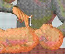
Fig. 237. Indirect cardiac massage by the method of two fingers
Step 4 (Drugs) - start medications if needed.
Indications for the use of drugs: despite adequate ventilation by 100% oxygen and indirect cardiac massage for 30 seconds, the heart rate remains 60 – 80 for 1 minute. A special solution of adrenalin should be prepared immediately for infant: 0. 1 ml of a 0. 1% solution of adrenalin in combination with 0. 9 ml of 0. 9% solution of natrium chloridum. 0. 2 ml/kg of body weight of this mixed solution should be injected intravenously for treatment of infant in case of bradycardia with 100 beats per minute. This injection may be repeated in 5 minutes intravenously or endotracheally. In case of significant bradycardia (less than 80-60 beats/minute), intracardiac injection of adrenalin should be done immediately.
Every 20 seconds 3 signs (natural respiration, heart rate, colour of the skin) should be evaluated to determine the effect of treatment.
After the occurrence of natural respiration and normalizing heart rate, oxygen inhalation should be continued until the skin becomes pink.
Infections of Newborn
The fetus normally synthesizes only very small amounts of immunoglobulins. However, after the twentieth week of gestation the intrauterine infections such as syphilis, rubella, cytomegalovirus infection and toxoplasmosis will stimulate the production of fetal IgM and IgA. The presence of a raised level of non-specific IgM antibody in the baby therefore suggests some kind of prenatal infection, at term baby cord levels of IgG are similar to those of maternal blood thus passively transferred antibody gradually disappears over the first few months of life. In " preterm” babies (of 32 weeks and less) placental transfer is not so efficient and their serum levels of IgG are low for the first three months of life. In a newborn baby phagocytosis is less efficient than normal and this contributes to the increased susceptibility to bacterial infection. Breast feeding confers some immunological advantages, particularly the transfer of IgA.
Sources of Infection
During pregnancy such organisms as rubeola virus and cytomegalovirus reach the fetus from the mother through the placenta. During birth the baby may be directly infected with organisms present in the birth canal and these include gonococci, group В streptococci and E. coli. After birth the infections with staphylococci, streptococci and blue pus bacillus (Pseudomonas aeruginosa) are derived from the environment.
Colonization of umbilicus, nose, throat and rectum takes place over the first few days of life. The organisms found in these sites are usually Gram-negative (E. coli and Ps. aeruginosa) in low-birth-weight infants, and Gram-positive (streptococci and staphylococci) in normal full term infants.
Clinical Features
The presenting symptoms and signs of neonatal infection are usually non-specific, especially in low-birth-weight babies or those who are already ill for other reasons. Lethargy and reluctance to feed are the first and the only indication of infection. Fever may be present but the temperature may be normal or even subnormal; the baby's colour is unnatural, it may be cyanosed or pale or even jaundiced. The liver and spleen are often enlarged. The infection is probably advanced. There may be fits and a bulging fontanelle in meningitis and rapid grunting respirations in pneumonia.
Laboratory Investigations
The object of investigation is to confirm the presence of infection, to localize its site and to determine the identity and sensitivities of the infecting organism. Blood, swabs and other material should be cultured. White cell counts may be helpful if the total count is about 7, 000 per cm3 after the fourth day of life with a marked shift left in the neutrophils. Serum levels of IgM are raised in the newborn following intrauterine infections so that a risen level after birth does not provide evidence of a postnatal infection.
Common Infections
Skin Infections
Most skin infections in the newborn are caused by staphylococci. The usual lesions are small pustules or a mild paronychia. Occasionally the skin around the umbilicus may become red and edematous indicating a more virulent umbilical stump infection than usual. In mild skin infections swabs should be taken from the infected areas which can then be painted with crystal violent paint of 0. 2 percent. More severe infections might lead to staphylococcal septicemia, pneumonia or osteomyelitis and should be treated with an appropriate systemic antibiotic such as cefazolin.
Thrush
Monilia infection is usually mild and affects the mouth and perianal skin. In the mouth there are small white plaques on the inside of the cheek or on the tongue which look like curds of milk but do not wipe off, the fungus passes down the alimentary tract and sets up a mild perianal skin infection which appears as a uniform erythema. In more severe cases, which are rare, the lung may be involved. Thrush is treated with nystatin mixture, 100 000 units in 1 ml four times a day after feeds. Nystatin cream aids the perianal skin.
Gastroenteritis
Fortunately this is now an uncommon infection in the newborn but when it occurs, it may give rise to a nursery epidemic. It is therefore wise to take particular notice of newborns that develop diarrhea. The pathogenic organism may be colibacillus (E. coli) or a virus or one of the salmonella group. As at gastroenteritis in another child, dehydration is the main problem and must be rapidly corrected. Antibiotic therapy should be used too.
Urinary Tract Infections
The diagnosis of urinary tract infection can be made only on revealing a significant bacteriuria and pyuria on a fresh clean specimen of urine. The baby is held out with its legs apart, the skin of the perineal area is stroked to stimulate micturition and the voided urine (after the first drops) is collected in a sterile container. In cases of urgency urine can usually be obtained by suprapubic bladder aspiration. Leucocyte counts of over 10 per cm3 and bacterial counts of more than 105 per ml on two consecutive specimens establish the diagnosis. The usual organism is E. coli. Although most of the babies with urinary tract infection will have a mild illness, some will be seriously ill with septicemia and jaundice. Appropriate antibiotics should be given and follow up should be arranged to decide on the need for radiological investigation of the urinary tract.
Pneumonia
Certain adverse factors during labor and delivery predispose to neonatal pneumonia, for example difficult delivery, infected liquor and aspiration of meconium. Gram-negative organisms are predominant. The baby appears ill and may have few respiratory symptoms. There may be recession of the chest wall, cyanosis, and tachypnea. X-rays should be taken to establish the diagnosis.
The meconium aspiration syndrome is caused by aspiration of meconium stained liquor during birth and is associated with asphyxia and post-term delivery. There are patchy areas of airway, obstruction and atelectasis and infection may be superimposed. The clinical picture may be difficult to distinguish at early onset of pneumonia but the radiological features of meconium aspiration are characteristic.
Meningitis
The disease is about four times commoner in pre-term than in full-term babies. The organisms are usually Gram-negative; E. coli and Ps. aeruginosa are the most common. The onset is usually in the first week and often on the first day, gradual with fever, poor feeding and vomiting as the commonest symptoms. The classical signs of meningitis are usually absent and lumbar puncture should be performed early in sick babies who have no obvious cause for their illness. Blood culture should be carried out to help in identifying the organism. Antibiotic therapy should be started as soon as possible, the choice of antibiotic depending on the obtained information about the organism.
Infections Acquired in Uterus
There is a surprising similarity in the clinical features of a group of non-bacterial infections acquired by the fetus during pregnancy and those presented in the first week or so after birth. Not all affected babies show all these features. The organisms concerned are: rubella virus, cytomegalovirus, toxoplasma gondii, herpesvirus hominis, treponema pallidum.
The clinical features in common are: central nervous system abnormalities, choroidoretinitis, osteitis, hepatosplenomegaly, anemia, purpura, intrauterine growth retardation.
Congenital Rubella
In addition to the clinical features listed above infants exposed to intrauterine rubella infection in the first three months of pregnancy may have eye defects, cardiac defects and damage to the eighth cranial nerves. The involvement of the central nervous system may lead to retarded mental development.
The diagnosis of congenital rubella infection may be confirmed by the findings of specific fluorescent antibody in the baby's struma. If both mother and baby have a raised rubella antibody the baby's titre must be measured again four to six months later. If it is still raised a retrospective diagnosis of congenital rubella can be made. Affected babies may excrete the virus for up to one year after birth and can be a source of infection to others.
Congenital Cytomegalovirus Infection
The fact that this infection occurs commonly is of importance. In the newborn it is usually symptomless, sometimes it gives rise to relatively mild effects and infequently causes a severe acute syndrome. The clinical infection may result in mental retardation in babies and there is some information to suggest that a significant number of mentally retarded children may have a subclinical infection with cytomegalovirus in the neonatal period. The finding of virus-specific IgM or the isolation of the virus indicates infection, but not necessarily a clinical disease. Retrospective diagnosis is made by finding a high titre of complement fixing antibody at birth and four to six months later.
Congenital Toxoplasmosis
The toxoplasma protozoon is commonly found in animals including the domestic ones which is a potent source of infection in man. During pregnancy the organism may be transmitted to the fetus from a mother with unrecognized mild infection. Most of the exposed infants are unaffected but the effect on the eyes and the brain in those that develop symptoms may be very severe. The diagnosis is established either by finding the organism in the CSF or by appropriate serological tests in mother and infant.
Antibiotics in Neonatal Infections
It is commonly considered unnecessary or unwise to use antibiotics for prophylaxis in the newborn. However it may be necessary to use them for a sick newborn before the laboratory confirmation of the nature of infection is made. In such circumstances all material for bacteriological diagnosis must be obtained before starting treatment.
The choice of antibiotic for a baby with a presumed infection depends on the probable site of infection and the knowledge of usual colonizing organisms in the particular hospital environment. Since these considerations will probably indicate the possibility of either a Gram-negative or Gram-positive organism it is usual to choose a broad spectrum combination of antibiotics such as ampicillin and cloxacillin, kanamycin and benzyl penicillin or gentamycin and cephazolin. In any case the antibiotic therapy must be reviewed if and when the organim and its sensitives are known. In case of meningitis chloramphenicol alone is an additional choice because of its wide spectrum and high concentration in the cerebrospinal fluid. The dose should not exceed 25 mg/kg/d in the first week of life, as high doses in preterm infants have produced collapse and death (“grey syndrome”).
Ophthalmia Neonatorum
It is a preventable disease occurring in newborn children due to maternal infection acquired as a result of carelessness at the time of birth; it used to be responsible for 50 per cent of blindness in children. Previously the disease used to be very severe but nowadays it is mild in nature.
There occurs dense infiltration of the bulbar conjunctiva, and the lids are swollen and tense. Later the lids become softer and more easily everted, the conjunctiva becomes puckered and velvety, and the blood stasis gives place to intense itching and congestion, with the free discharge of pus, serum and often blood. In some cases a false membrane forms, so that the case resembles a membranous conjunctivitis.
There is a great risk of corneal ulceration in untreated gonococcal ophthalmia neonatorum, since this organism has the power of invading intact epithelium. The slightest haziness should be viewed with apprehension, hardly metastatic stomatitis and arthritis occur. The arthritic manifestations usually appear in the third or fourth week and affect the knee, wrist, ankle or sometime elbow.
In inadequate cases serious sequelae may occur. If the corneal ulceraton heals without perforation there is always much scarring of this tissue, but the nebula clears more in babies. Perforation may be followed by anterior synechial, adherent leucoma, partial or total anterior staphyloma, anterior capsular cataract or panophthalmitis. When vision is not completely destroyed but is seriously impaired by the corneal opacities, the development of macular fixation which takes place during the first six weeks of life is impaired, resulting in the development of nystagmus which persists throughout life; this may not manifest until a later date.
The inclusion conjunctivitis is caused by a virus.
As the disease is preventable, so a prophylactic treatment is, therefore, of prime importance. Any suspicious vaginal discharge during the antenatal period should be treated. The newborn baby's closed lids should be thoroughly cleaned and dried. A drop of silver nitrate solution, one percent, should be instilled into each eye. The eyes must be carefully watched during the first week.
If the disease is established and there is any purulent discharge, the eyes must be irrigated with warm saline and intensive therapy with penicillin started, using drops in a concentration of 5, 000 units per ml every minute, for half an hour. Repeated irrigations are unnecessary since, in the first place, penicillin remains effective in the presence of pus and, in the second, the discharge rapidly disappears. Any pus that does accumulates is wiped away with moist pledgets of cotton-wool, Penicillin drops continued at five minute interval for a further half-hour, and the treatment is consolidated by half hourly and then hourly instillations for two days or so. Astringent lotions are then employed.
An alternative is a systemic administration of full course of sulphonamides as sulphadiazine.
In default of treatment by antibiotics, or when dealing with insensitive organisms, reliance must perforce be placed on the old-fashioned treatment of repeated application of silver nitrate together with repeated irrigations with antiseptic solution. Such treatment, however, is most unsatisfactory: corneal complications frequently supervene and recovery is often slow.
Atropine should be used in all cases in which the cornea is involved since this is always accompanied by some iritis; corneal complications require very active treatment.
Birth Injuries
Birth injuries are those injuries which occur during the birth of the child. These injuries were described in special chapter.
Care of the Neonate
The care of the neonate should provide conditions favouring its development. The main principle underlying the care of the neonate is its protection from unfavourable effects of the environment, especially from possible infection.
Specially trained nurses are admitted to take care of the neonate. An obstetrician takes care of the neonate at small hospitals. Auxiliary personnel are not admitted to contact the neonate.
At the beginning of the shift, the medical personnel should obligatory wash their hands (to the elbows) using soap and a brush and then disinfect them (alcohol, lysoform, chloramine solutions, etc. ). The nurse should wash her hands and disinfect them each time before swaddling the infant.
When the infant is brought into the neonate room from the delivery room, it is wrapped in warmed-up cloths, placed in bed and its condition is observed thoroughly. It should be remembered that a neonate may vomit or develop cyanosis; the umbilical stump may bleed.
Two times a day (before the first feeding and in the evening) the neonate should be given a hygienic treatment. During this procedure the infant is given a general outer examination, all skin folds are inspected, the bandage on the umbilical stump is checked, and the prescribed procedures are done. The weight and temperature of the infant are also measured.
Proper care of the infant's skin is very important since abrasion and intertrigo may become the site of entrance of infection. Purulent diseases of the skin may become the cause of sepsis.
The body of the infant is inspected during the morning and evening toilet; special attention should be paid to the skin folds where intertrigo would most likely occur (behind the ears, on the neck, in the armpits and groins, etc. ).
The infant should not take bath before the umbilical cord falls off since infection is likely to enter through the non-healed umbilical wound. The washing therefore consists only in wiping the face with a piece of cotton wool soaked in warm boiled water or a 2 per cent solution of boric acid. The ears, the skin behind the ears, the skin folds on the neck, the palms and soles should be wiped with a wet cotton wool pad. The body should then be dried up.
If any reddening appears on the skin, the affected sites should be coated with sterile vaseline or sunflower oil.
The neonate should be swaddled anew after each urination or defecation (in the intervals between nursing). Soiled swaddles are replaced by clean ones, while the lower part of the infant's body should be washed in a jet of warm water. In doing so it is necessary to protect the umbilical region from wetting with water. The skin should then be dried up by touching (not rubbing! ) it with a clean cloth readily absorbing moisture. For prophylactic purposes the skin of the buttocks and the groins should be coated with sterile oil.
Intertrigo would normally be the result of inadequate care of the neonate. Treatment of intertrigo includes improved care, prevention of the skin from soiling with faeces or urine. If intertrigo is insignificant (skin is only reddened), application of sterile oil would usually be enough. If the reddened skin is macerated, a 3 per cent solution of potassium permanganate should be used to treat the affected skin with subsequent powdering with the preparation of the following composition: boric acid 1. 0 g, bismuth subnitrate, zinc oxide 5. 0 g of each, talcum and starch (amylum), 50. 0 g of each.
Prophylaxis of pyodermitis is especially important. If any purulent foci appear on the skin, the infant should be isolated from other neonates. Each pustule should be treated with an alcoholic solution of gramicidin (0. 04 per cent), or potassium permanganate (3 per cent). Penicillin is often prescribed intramuscularly. The neonate should be under constant medical observation.
Care of the sense organs. The eyes of the neonate should be treated with cotton wool soaked in a 2 per cent solution of boric acid. Each eye is treated with a separate ball of cotton wool, moving it from the outer canthus of the eye to the inner one.
Wiping the ears with wet cotton during the morning toilet will normally be enough; it is unnecessary to clean the meatus.
The nostrils should only be treated if the respiration is difficult and much mucus or crusts are accumulated in them. The openings are then wiped carefully with a piece of cotton wool twisted into a cord and soaked in vaseline oil.
It is prohibited to wipe the mouth mucosa since it can easily be damaged. Even in case of thrush (the result of inadequate care and feeding) the mucosa should not be wiped but only coated carefully with a 10 per cent solution of borax and glycerol.
Self Test
1. Which of the following should be used for evaluation of the status of neonate with asphyxia?
A. spontaneous respiration, heart rate, colour of the skin
B. Apgar’s scale
2. Adrenalin solution should be injected intravenously for treatment of infant in case of:
A. bradycardia 100 beats/minute
B. bradycardia 60-80 beats/minute
C. absence of heart rate
3. In case of absence of heart rate adrenalin may be introduced
A. intravenously.
B. intracardially.
C. intratracheally.
4. The eyes of neonate should be treated with cotton wool soaked in
A. 2 per cent solution of boric acid.
B. 2 per cent solution of iodine.
C. camomile tea.
D. 3 per cent solution of hydrogen peroxide.
5. Clinical signs of ophthalmia neonatorum are all of following except for:
A. intense itching and congestion
B. discharge of pus, serum or blood
C. nonclosed eyes
CHAPTER 38. EMERGENCY OBSTETRIC CARE
Shock in Obstetrical Practice
Shock is a state of circulatory failure characterized by inadequate tissue perfusion. Blood flow is insufficient to provide the nutritional requirements of cells and remove the waste products of metabolism. This insufficiency leads to cellular dysfunction and, ultimately, death. Correct management of patients in shock involves treating both the underlying cause of shock and the physiologic abnormalities associated with the shock.
Classification
The causes of shock can be classified on the basis of 4 major pathologic mechanisms involved.
· Hypovolemic shock. The chief abnormality in hypovolemic shock is decreased intravascular volume, which may occur as a result of loss of blood or plasma, or fluid and electrolytes. These losses may be exogenous or endogenous (“third-spacing” hematomas).
· Cardiogenic shock. The chief abnormality in cardiogenic shock is abnormal cardiac function due to arrhythmia, “pump failure” or valvular dysfunction.
· Obstructive shock. The chief abnormality in obstructive shock is an impediment to filling of the right or left ventricle (decreased preload). If decreased filling is sufficiently severe, the resulting fall in cardiac output causes shock. Obstruction may occur in the systemic circulation (obstruction of the vena cava) or pulmonary circulation (massive pulmonary embolus) or may be due to pericardial disease (cardiac tamponade) or cardiac disease (atrial myxoma).
· Distributive shock. The chief abnormality in distributive shock is abnormal distribution of vascular volume due to changes in vascular resistance or permeability. The end result is a decreased ventricular filling that leads to inadequate cardiac output. The derangement of vascular volume characterizing distributive shock may occur as a result of sepsis, anaphylaxis, or neurogenic shock.
The classification of shock according to mechanism and common causes is given in Table 26.
Table 26
Classification of shock by mechanism and common causes
| Hypovolemic shock Loss of blood (hemorrhagic shock) External hemorrhage: Trauma Gastrointestinal tract bleeding Vaginal (uterine) bleeding Internal hemorrhage: Hematoma Hemothorax Hemoperitoneum Loss of plasma Burns Exfoliative dermatitis Loss of fluid and electrolytes External Vomiting Diarrhea Excessive sweating Hyperosmolar states (diabetic ketoacidosis, hyperosmolar nonketotic coma) Internal (“third spacing”) Pancreatitis Ascites Bowel obstruction Cardiogenic shock Dysrhythmia Tachyarrhythmia Bradyarrhythmia “Pump failure” (secondary to myocardial infarction or other cardiomyopathy) Acute valvular dysfunction (especially regurgirtant lesions) Rupture of ventricular septum or free ventricular wall Obstructive shock Tension pneumothorax Pericardial disease Disease of pulmonary vasculature (massive pulmonary emboli, pulmonary hypertension) Cardiac tumor (atrial myxoma) Left atrial mural thrombus Obstructive valvular disease (aortic or mitral stenosis) Distributive shock Septic shock Anaphylactic shock Neurogenic shock Vasodilator drugs Acute adrenal insufficiency |
Any types of shock may be found in obstetrical practice, but the most common ones are hemorrhage shock and distributive shock (septic, anaphylactic).
Diagnosis
A. A suspect shock in the following signs is present:
1. Hypotension. Systolic blood pressure of 90 mm Hg or less in adults usually signifies hypotension. Some healthy adults may have systolic blood pressure that is normally this low; conversely, patients with preexisting hypertension may develop shock at blood pressure levels within the normal range. Orthostatic vital signs should be measured if equivocal blood pressure readings are found and no spinal injury exists.
2. Orthostatic change in vital signs. Orthostatic vital signs should be measured in patients who are not clearly hypotensive in the supine position. Both blood pressure and pulse are measured in the supine and then in the sitting (legs dangling over the side of the bed) position. If no change occurs in the sitting position, repeat the determination with the patient standing. Wait about 3-5 minutes between measurements to allow pulse and blood pressure to stabilize. A drop in systolic blood pressure of 10-20 mm Hg or more associated with an increase in pulse rate of more than 15 beats/min suggests depletion of intravascular volume. An increased heart rate with no change in blood pressure in the seated or erect position may be seen in some patients with mild hypovoilemia.
On the other hand, normovolemic patients with autonomic neuropathies or those taking certain medications (some antihypertensive drugs) may demonstrate an orthostatic fall in blood pressure, usually without an associated increase in pulse rate.
3. Tachycardia. Tachycardia, although a non-specific finding, is usually present in mild to moderate shock. An orthostatic fall in blood pressure will help confirm the cause of tachycardia.
4. Adrenergic responses. Restlessness, anxiety, and diaphoresis may accompany the shock state.
5. Peripheral hypoperfusion. Cool or mottled extremities (livedo reticularis) and weak or impalpable peripheral pulses are signs of peripheral hypoperfusion.
6. Altered mental status. Patients in shock may demonstrate normal mental status or may be restless, agitated, confused, lethargic, or comatose as a result of inadequate perfusion of the brain.
B. Determining severity of shock.
1. Mild shock. Mild shock is defined as decreased perfusion of nonvital organs and tissue only (skin, fat, skeletal muscle, bone). These tissues can survive relatively long periods of decreased perfusion without undergoing irreversible changes. Mentation is unimpaired, urine output is normal or only slightly decreased, and metabolic acidosis is absent or mild.
2. Moderate shock. Moderate shock is defined as decreased perfusion of vital organs other than heart and brain (liver, kidneys, and others). These organs do not tolerate hypoperfusion as long as fat, skin, and muscle do. Oliguria (urine output, 0. 5 ml/kg/h in adults) and metabolic acidosis are present, but the sensorium is relatively intact.
3. Severe shock. Severe shock is defined as inadequate perfusion of the heart or brain. The compensatory mechanisms of shock act to preserve blood flow to these 2 vital organs at the expense of all others. Thus, in advanced shock there is constriction of all other vascular beds. In addition to severe oliguria and acidosis, altered mentation and signs of cardiac hypoxia (abnormal ECG, decreased cardiac output) occur.
Initial Treatment:
General Measures
1. Position the patient: Place the patient supine and level to maximize blood flow to the brain. (Remember to prevent unnecessary heat loss with a blanket when the patient is not being examined).
2. Establish adequate oxygenation: Open or maintain the airway. Support inadequate ventilation with a bag-mask combination or endotracheal tube. Obtain arterial blood gas measurements as a guide both to the adequacy of oxygenation and to the severity of hypoperfusion, as indicated by the presence or absence of metabolic acidosis. Pending the results of blood gas measurements, give oxygen, 5-10 l/min, by mask or nasal prongs.
3. Stop obvious external hemorrhage: Use direct manual compression. Blind clamping of vessels should be avoided, since it causes further injury. Tourniquets are rarely indicated except in traumatic amputation.
4. Gain intravenous access: The type and number of venous access sites, as well as the type and size of the intravenous catheters, depend in part on the type of shock suspected.
a) Suspected hypovolemic shock. In patients with apparent hypovolemic shock, insert 2 large-bore
|
|
|
