 |


Caldwell-Moloy classification. Diagnosis of contracted pelvis.
|
|
|
|
Caldwell-Moloy classification
Caldwell-Moloy classification (1933) are widely used abroad. It is based on structural features of the pelvis (Fig. 194):
· gynecoid (female type of pelvis)
· android (male pattern)
· anthropoid (inherent in primates)
· platipelloid (flat)
Gynecoid pelvis - the normal female pelvis: a rounded oval pelvis with well-rounded anterior and posterior segments.
The main features are:
• A spacious and well-rounded posterior segment
• An inlet with a slightly ovoid or round shape
• A wide, well-rounded (anterior segment)
• A sacrosciatic notch of medium size
• An average sacral inclination and curvature
• A wide subpubic arch
• Wide interspinous and intertuberous diameters
• Bones ranging from medium to delicate in structure
• Bones ranging from medium to heavy in structure
• Engagement in this type of pelvis occurs most frequently with the fetus in a transverse position, followed in frequency by the posterior and anterior positions. The clinician should be alerted by this type of pelvis that the possibility of posterior positions exists.
Android pelvis is a pelvis with a wedge-shaped inlet and narrow anterior segment; used to describe a female pelvis with characteristics usually found in the male. The main features are:
• A wedge-shaped inlet
• A narrow retropubic angle (anterior segment)
• A flat, wide posterior segment
• A narrow sacrosciatic notch
• A forward sacral inclination
• A narrow wedge-shaped “Gothic” subpubic arch
• Converging side walls, narrow interspinous and intertuberous diameters
• Bones ranging from medium to heavy in structure
Anthropoid pelvis is a female pelvis in which the anteroposterior diameter of the inlet equals or exceeds the transverse diameter. The main features are:
· A long, narrow, oval-shaped inlet
· A long, narrow, well-rounded anterior segment
· A long, narrow posterior segment
· A very wide, shallow sacrosciatic notch
· A long, narrow sacrum with average inclination and curvature
· A slightly narrow subpubic arch
· Straight side walls with below-average interspinous and intertuberous diameters
· Medium to delicate bones
Engagement in this type of pelvis occurs with the fetus in either an anterior or transverse position, but the anterior position appears to be more characteristic.
Platypellic pelvis (platypelloid pelvis)is a pelvis which is shortened in the anteroposterior aspect, with a flattened transverse oval shape. The main features are:
· A transverse, oval-shaped inlet
· A very wide, round retropubic angle
· A very wide, flat posterior segment
· A narrow sacrosciatic notch
· Average sacral inclination
· A very wide subpubic arch
· Straight side walls with very wide interspinous and intertuberous diameters
· Bones ranging from medium to delicate in structure
Engagement in this type of pelvis will almost always occur with the fetus in a transverse position. Because of the flatness of the shape of this pelvis, the internal rotation of the vertex canbe limited, causing a deep transverse arrest.
|
|
|

Fig. 194. Pelvis types by Caldwell-Moloy classification
Diagnosis of contracted pelvis.
The diagnosis of anatomically contracted pelvis is of importance to prevent obstetrical complications during delivery.
The diagnosis is based on:
· Anamnesis vitae: conditions of growing up in childhood, infectious diseases, rachitis, etc. Conditions of life, nutrition, medical diseases, traumas, if any, etc.
· Previous history. One should pay attention to the character of previous delivery, if any. The duration of labor, character of labor pains, baby’s body weight and general condition just after birth.
· Examination of woman: constitution, height, body weight, size of footwear. Short women with signs of infantilism, rachitic changes in skeleton, ankylosis and dislocations of the hip joint may have contracted pelvis. The shape of Michaelis’ rhomboid is very important for the diagnosis. Two sizes are distinguished in the rhomboid: longitudinal (between its top and bottom corners) and transverse (between lateral corners). Each of these sizes is 11cm, thus the rhomboid is regular in the normal pelvis (Fig. 195). In anatomically contracted pelvis it is irregular. The shape of the abdomen is also changed in marked contracture of the pelvis. The growing uterus fails to be held inside the abdominal cavity (which is shorter in short women) and deviates upwards and anteriorly. An acuminate abdomen is formed in primigravida in whom the abdominal wall is resilient while in multiparous women the abdomen becomes pendulous (Fig. 196, 197). The fetal head fails to enter a contracted pelvis at the end of pregnancy and floats high above the inlet till onset of labor.
· Pelvic measurement means external pelvimetry (interspinous diameter, intercristal diameter, intertrochanteric diameter, external conjugate). The wrist girth is measured to understand the thickness of the pelvic bones, or internal volume of the pelvic cavity which depends on the bone thickness. The circumference of the wrist girth, named Soloviev’s index, is normally about 14-15 cm. The more the circumference is, the less the internal volume of the pelvic cavity is.
· Internal pelvimetry may be done with the help of ultrasound measurement of internal diameters of the pelvic cavity, X-ray pelvimetry (rare, and only in non-pregnants).
· External obstetric examination: the lying of the fetus (transverse and oblique), breech presentations, extensive head presentations are frequent in patients with narrow pelvis.
· Vaginal examination usually helps in diagnosis of pelvic contracture: diagonal conjugate, palpation of the sacral promontory (if it can be reached by the examining finger), deformation of the pelvic cavity (if any) because of the tumor, etc.
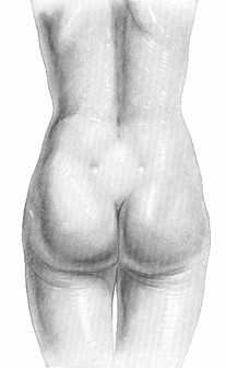
Fig. 195. Michaelis’ rhomboid is regular with the normal pelvis
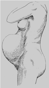
Fig. 196. An acuminate abdomen is characteristic for primipara with narrow pelvis

Fig. 197. Pendulous abdomen is characteristic for multipara.
Justo Minor Pelvis
Justo minor pelvis has the same shape as a normal one while all diameters are shortened. For example, typical sizes are: interspinous diameter – 24 cm, intercristal diameter – 26 cm, intertrochanteric diameter – 29 cm, external conjugate – 18 cm, etc. (Fig. 198).
|
|
|
The rhomboid is shortened both in vertical and horizontal axis.

Fig. 198. Justo-minor pelvis
Mechanism of Labor in Justo Minor Pelvis
The 1st moment is a strong flexion of the head. The sagittal suture aligns with one of oblique diameters of the pelvic inlet. The biparietal diameter of the head passes the oblique diameter of the pelvis which is longer than the anteroposterior one. The engaging diameter of the head is a small oblique (suboccipitobregmatic) diameter. The leading point (denominator) is a small (posterior) fontanelle. A strongly flexed head descends into the pelvic cavity and then performs the same movements as in normal mechanism of labor: the internal rotation, extension, external restitution. Because of narrow pubic angle, the posterior cranial fossa comes in direct contact with the symphysis as the head passes the pelvic outlet plane. The head is therefore displaced toward the perineum to a greater extent than in normal pelvis, the perineal tissues undergo greater extension and deep laceration of the perineum may occur. But all movements are comparatively slow and require much effort of the parturient.
The head of the delivered fetus is elongated in the direction of the occiput (dolichocephalic shape) and a considerable swelling is formed in the region of the posterior fontanelle (Fig. 199).

Fig. 199. Dolicho-cephalic shape of the head.
Transverse Contracted Pelvis
One or more transverse diameters may be shortened in transverse contracted pelvis. Transverse contracted pelves are usually elongated in anteroposterior direction. There are a lot of varieties of transverse contracted pelvis, which depend on the shortened diameter, degree of shortening, etc. For example, sizes of transverse contracted pelvis are: interspinous diameter – 23 cm, intercristal diameter – 26 cm, intertrochanteric diameter – 28 cm, external conjugate – 21 cm. Diagonal and true conjugate may also be normal or even bigger than in normal pelvis (Fig. 200). The rhomboid of Michaelis is elongated in its vertical axis so that its upper and lower angles are acute, and lateral ones are obtuse.

Fig. 200. Transverse contracted pelvis
Mechanism of Labor in Transverse Contracted Pelvis
The 1st moment is flexion of the head. The sagittal suture aligns with one of oblique or anteroposterior diameter of the pelvic inlet (depending on shortening of transverse diameter), the engaging diameter of the head is suboccipitobregmatic (small oblique), the denominator (leading point) is a small (posterior) fontanelle. The head enters the pelvic cavity and sometimes even reaches the pelvic floor without any rotation. This condition is termed as high anteroposterior situation of the sagittal suture. Further the head is delivered as in occiput presentation.
Flat Pelves
Rachitic Flat Pelvis
This type of narrow pelvis is characterized by reduction of the anteroposterior diameter of the pelvic inlet. The external conjugate is shortened; the upper portion of lumbar rhomboid is reduced. As for internal constitution (structure) of the pelvis, it is caused by special changes of the sacrum: the sacrum is slightly flattened; the corpus and the apex of the sacrum are usually deviated backward, while the base of the sacrum is moved up forward. That’s why the anteroposterior diameter of the pelvic inlet is reduced, while anteroposterior diameters of other planes are enlarged, although they may be normal in sizes. On vaginal examination one can find protruding sacral promontory, which can be easily reached by an examiner’s finger. Exostoses are common in rachitic pelvis.
|
|
|
Typical sizes of rachitic flat pelvis are: interspinous diameter – 28 cm, intercristal diameter – 28 cm, intertrochanteric diameter – 30 cm, external conjugate –17 cm (less than 20 cm), diagonal conjugate, true conjugate are less than normal (Fig. 201).

a. b.
Fig. 201. Rachitic flat pelvis: a - front view; b - side view.
Mechanism of labor in rachitic flat pelvis is changed because of the shortened anteroposterior diameter of the pelvic inlet.
The 1st moment is when the sagittal suture aligns with transverse diameter of the pelvic inlet for a long time. Since the anteroposterior diameter is shortened, the head fails to engage and preserves this position for a few hours.
Moderate deflexion of the vertex occurs during this moment, as a result of which the large (anterior) fontanelle becomes a leading point (denominator), and bitemporal diameter of the head becomes the engaging diameter. The typical sign of this mechanism is anterior asynclitism, which occurs more frequently than posterior asynclitism. In anterior asynclitism the posterior parietal bone rests against anteriorly displaced promontory where it is retained, while the anterior parietal bone descends gradually into the pelvis (Fig. 202).

Fig. 202. Anterior asynclitism in flat pelvis
The sagittal suture is now situated nearer the promontory (Fig. 203).

Fig. 203. Sagittal suture is near the promontory.
In this position (with the sagittal suture aligned with the transverse diameter, closer to the promontory, and anterior fontanelle below the posterior one) the head remains engaged until it is sufficiently molded, after which the posterior parietal bone slips off the promontory and the asynclitism disappears. The next moments are the same as in normal pelvis.
The 2nd moment is internal rotation of the head, with occiput turned to the symphysis pubis, and face turned to the promontory. The 3rd moment is deflexion of the head, the 4th is internal rotation of the shoulders and external rotation of the head, the 5th moment is lateral flexion of the trunk and shoulders delivery, the 6th moment is expulsion of the trunk.
After the head has passed the contracted inlet plane of the pelvis, the expulsion of the fetus becomes very rapid because the pelvic cavity is normal or enlarged, while the pelvic outlet is wider than normal. Complications are: fetal hypoxya due to precipitate delivery, intracranial injuries of the fetus, injuries of the perineum and other soft tissues of the birth canal.
Simple Flat Pelvis
Simple flat (platypellic) pelvis is characterized by reduction of all anteroposterior diameters of the pelvis. This occurs due to displacement of the sacrum: it is pushed in the pelvic cavity towards the symphysis pubis. Thus all anteroposterior diameters of the pelvic planes are less than in the normal pelvis. In a simple flat pelvis the Michaelis’s rhomboid is shortened in the vertical direction so that its upper and lower angles are obtuse, while the lateral ones are acute.
Typical sizes of simple flat pelvis are: interspinous diameter – 26 cm, intercristal diameter – 28 cm, intertrochanteric diameter – 30 cm, external conjugate – less than 20 cm, diagonal and true conjugates are less than in normal pelvis (Fig. 204).
Mechanism of Labor in Simple Flat Pelvis
The head enters the pelvis in the same way as in flat rachitic pelvis: the sagittal suture aligns with the transverse diameter of the pelvic inlet, the engaging diameter of the fetal head is bitemporal diameter, and the leading point (the denominator) is anterior (large) fontanelle. Further it descends into the pelvic cavity and is delivered as in the occiput presentation. But sometimes the head fails to perform the internal rotation because the anteroposterior diameters of the midpelvic and outlet pelvic planes are also reduced. The head enters the pelvic cavity and sometimes even reaches the pelvic floor, while the sagittal suture remains aligned with the transverse diameter. This condition is termed as lower transverse situation of the sagittal suture. In some cases the head rotates with its occiput anteriorly as it reaches the pelvic floor and labor then ends spontaneously. The rotation of the head on the pelvic floor is usually complicated by lacerations of the perineum. The duration of labor in simple flat pelvis is marketly longer than in rachitic pelvis, but more preferable for the fetus. Typical complications are: secondary uterine inertia, fetal hypoxia, maternal lacerations.
|
|
|
The presenting parietal bone of the delivered head bears a large swelling: the shape of the head may be oblique, named a brachicephalic shape (Fig. 205).

Fig. 204. Simple flat pelvis

Fig. 205. Brachicephalic shape of the head
Flat pelvis with reduction of anteroposterior diameter of the 2nd plane of the pelvis
Anteroposterior diameter of the second plane of the pelvis is the only shortened size in this pelvis. This type of pelvis is a result of failed development of skeleton at the age of 12-15 years due to hyperandrogenic and hypoestrogenic condition. There is absence of the sacral curvature, the sacrum is significantly flat, thus the anteroposterior diameter of the 2nd plane is shortened. The diagnosis of this pelvis is rather difficult. The external diameters of the pelvis are not changed. The general constitution is normal. There are no marked diseases in the anamnesis. An experienced obstetrician may pay attention to the flat sacrum during vaginal examination. Sonography measurement of internal pelvic sizes helps to make the diagnosis.
Mechanism of labor at this pelvis is not well studied. The 1st moment is the same as in normal pelvis. But the 2nd moment (internal rotation of the head) is usually failed due to shortened anteroposterior diameter; cephalo-pelvic disproportion is often present in this pelvis.
Generally Contracted Flat Pelvis
This shape of pelvis is characterized by shortening of all diameters of the pelvis with prevalence of anteroposterior diameters (Fig. 206). The mechanism of labor is usually similar to that at other flat pelves, but then the engagement of the head fails. Labor at generally contracted pelvis is very difficult.

Fig. 206. Generally contracted flat pelvis.
Effect of Contracted Pelvis on Pregnancy and Labor
The general course of pregnancy is not much affected. However, the following may occur: malpresentations may happen 3-4 times more often and incidence of unstable lie, premature rupture of amniotic membranes is also increased.
The course of labor is greatly modified depending on degree of pelvic contraction and presentation of the fetus. There is an increased incidence of early rupture of membranes. The incidence of cord prolapse is also higher. There is a marked tendency of prolonged labor due to secondary uterine inertia, and increased incidence of operative labor. The injuries of maternal tissues may occur spontaneously or depend on operative delivery. There may be lacerations of the perineum, cervix, vagina, failure of the pelvic junctions (symphysitis), and rupture of the uterus. Fetal risks are due to trauma and asphyxia. These may occur due to extreme molding of the head, prolapse of the cord, etc.
Cephalopelvic disproportion in labor
The influence of the contracted pelvis on the process of labor and delivery of the fetus lies in the development of so-called cephalo-pelvic disproportion. Cephalo-pelvic disproportion (CPD) is the disparity in the relation between the fetal skull and maternal pelvis dimensions during the process of labor. This leads to difficult labor or abnormally slow progress of labor. Other terms that are often used interchangeably with CPD are dystocia of labor, dysfunctional labor, failure to progress (lack of progressive cervical dilatation or lack of descent), obstructed labor with subsequent complications, such as fetal distress, injuries to the fetus, intrauterine fetal death, maternal injuries (lacerations of the cervix, perineum, rupture of the uterus, and others) and bleedings. All these complications are responsible for increasing of maternal and perinatal morbidity and mortality rates.
|
|
|
To define CPD, a definition of normal labor must be understood and accepted. Normal labor is defined as uterine contractions that result in progressive dilation and effacement of the cervix. The progress of labor as well as successful outcome is dependent on so-called 3 P’s. They are three main components that may influence the fetus’s ability to pass through the birth canal with relative ease:
|
|
|
