 |
Fig. 154. Classic podalic version with fully dilated cervix (internal version). The internal hand is introduced into the vagina for grasping the leg.
|
|
|
|
Identification of the leg
A leg lying near the abdominal wall of the mother should be found for version from the longitudinal presentation. The choice of the leg in transverse presentation depends on type of the fetus position: the inferior leg should be grasped if the fetus back is turned to the maternal abdominal wall. If the fetus back is turned to the maternal spine, the superior leg should be grasped.
To identify the leg, the hand feels the side of the fetus and slides from the armpit to the pelvic end, and further by the thigh to grasp the leg. The external hand should assist by moving the pelvic end down toward the internal hand of the operator (Fig. 155).

Fig. 155Internal version. Identification of the leg.
Grasping the leg
This can be done by two methods:
· The leg is grasped by the entire hand, i. e. four fingers are placed on the anterior part of the leg while the thumb is placed on the calf to reach the popliteal fossa (Fig. 156).
· The index and the middle fingers grasp the malleolus, while the thumb supports the sole. The former method is however more reliable (Fig. 157).
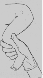
Fig. 156. Internal version. Grasping the leg: the leg is grasped by the entire hand.

Fig. 157. Internal version. Grasping of the leg: the leg is grasped by two fingers.
Version proper
After a firm grip is placed on the fetal leg, the external hand moves the fetal head upwards, i. e. to the fundus of the uterus. During the course of this manipulation, the internal hand pulls the leg outside the vagina. The version is considered complete when the leg is extracted from the pudendal cleft to the knee (Fig. 158). This indicates that the fetal assumes the longitudinal position. The arm may be prolapsed with the discharge of amniotic fluid in transverse presentation. The prolapsed arm should not be corrected but only pulled by a sterile gauze strip towards the symphysis. As the fetus is then turned, the arm retracts spontaneously.
Just after version the fetus should be extracted by the leg or podalic end.
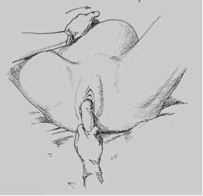
Fig. 158. Internal version. The leg is extracted from the pudendal cleft to the knee.
Extraction of the Fetus by Podalic End
Whenever complications endangering the mother and the fetus arise, labor in breech presentation should be ended by extraction of the fetus. This method is also used after the podalic version with a fully dilated cervix.
Extraction differs from manual assistance. Manual assistance in breech presentation is given after spontaneous delivery of the fetus to the lower angle of the shoulder-blades, its objective being to deliver the arms and the head.
Extraction of the fetus by the pelvic end is an operation in which the entire fetus, beginning with the legs, is extracted. The operation is performed by the hand only.
Indications:
· Severe diseases requiring urgent termination of labor (eclampsia, heart diseases, etc);
|
|
|
· Occurrence of asphyxia of the fetus;
· After the classical podalic version.
Requisite conditions:
· Fully dilated cervix;
· Rupture of the fetal membranes;
· Absence of cephalopelvic disproportion.
The fetus is extracted by one leg (in single footling) or by two legs (in double footling presentation).
Extraction by the leg or legs
If the presenting leg is in the vagina, it is extracted by two fingers. The leg is grasped in the following way: the thumb is placed on the calf, and the other four fingers — on the anterior surface of the leg. The leg may be grasped by both hands: both thumbs are then placed on the calves. This grip on the leg prevents its fracture (Fig. 159).
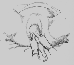
Fig. 159. Extraction by the leg: the leg is grasped by both hands.
The downward traction should then be applied to the leg. As the leg appears from the birth canal, the operator’s hand should move farther along the infant leg toward the pudendal cleft. The downward traction should be continued until the forthcoming buttock and the iliac bone are brought beneath the symphysis. The forthcoming thigh is then grasped by both hands and lifted slightly (Fig. 160).
The fatal body is thus flexed and the aftercoming buttock is delivered over the perineum.
No attempts should be taken to free the aftercoming leg for it may be fractured; the leg will be delivered spontaneously during further traction.
After the second buttock has been delivered, one hand should grasp the anterior thigh (the thumb being positioned on the sacrum), while the index finger of the other hand should engage the posterior groin of the fetus with the thumb resting on the sacrum (Fig. 161). The other fingers should be pressed against the palm. With the hands thus gripped on the buttocks, the operator continues the downward traction.
After the aftercoming leg is delivered, it is taken by the hand in the same manner and the fetus is extracted to the navel. The cord is checked for the absence of tension, and the fetus is extracted to the lower margin of the shoulder blades. Now the arm and the head are freed by manoeuvres as specified for manual assistance.
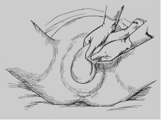
Fig. 160. Extraction by the leg: the forthcoming thigh is grasped by both hands and lifted slightly.
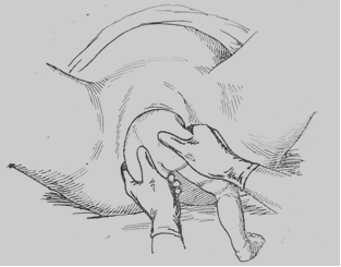
Fig. 161. Extraction by the leg: pulling the fetus till the navel.
Extraction by the Groin
Extraction of frank breech presentation begins with inversion of one index finger into the anterior fetal groin (Fig. 162). The traction is continued until the anterior buttock is freed and the iliac bone is brought beneath the pubic arch. Traction is only applied when the uterus contracts, in order to add to the traction effort. The other hand should assist by grasping the wrist of the operating hand (Fig. 163). When the anterior iliac bone is brought beneath the symphysis, the direction of traction should be changed to the upward one. The fetal torso is flexed and the posterior buttock is disengaged. The index finger of the other hand should now be inserted into the other groin. Traction is continued until the fetus is delivered to the lower margin of the shoulder blade. The legs are freed spontaneously.
The arms and the head are freed as in manual assistance.
Extraction by the groin is one of the most difficult obstetric operations. The operating hand is quickly fatigued, the buttocks move slowly. It is especially difficult to extract the fetus with a high position of its buttocks.
|
|
|
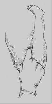
Fig. 162. Extraction by the groin.

Fig. 163. Traction by the groin.
Possible complications
The following complications may occur in extraction (manual assistance): spasm of the uterine cervix, extension of the arms, posterior presentation, etc.
In order to remove the spasm of the cervix os, the operation is performed with anesthesia. Spasms may be prevented by giving (before operation) 1 ml of a 1% papaverine solution and 1 ml of a 0. 1% atropine solution.
In order to correct the arm (when it is extended), four fingers should be inserted into the vagina and the arm pulled down along the face and chest (the fetus “washes” its face).
Extraction of the fetus is also difficult with persistent posterior position (the chin turns towards the symphysis and the occiput towards the sacrum). To disengage the head in posterior position, the following manipulations are required. The tip of the index finger is placed into the fetal mouth to flex the head, while the other hand of the operator should extract the fetus until the nose bridge (fulcrum) is brought beneath the symphysis. The torso of the fetus is then bent anteriorly (toward the mother’s abdomen) and the head is delivered over the perineum. If the head is extended and the finger fails to reach the mouth, the torso of the fetus should be flexed on the mother’s abdomen, while the other hand of the operator should grip the shoulders with the fingers (like a fork) to assist extraction of the fetus. The head passes the pelvis by its vertical diameter, the fulcrum being the region of the hyoid bone.
Self Test
1. The external version in transverse and oblique presentation should be done after
A. 35th week of pregnanсy.
B. 39th week of pregnancy.
C. 32nd week of pregnanacy.
D. the beginning of labor.
2. The external version in transverse and oblique presentation should be done
A. in antenatal clinic.
B. in hospital.
C. at home.
D. in operating hall.
3. The external version of the fetus should be done
A. without anesthesia.
B. with local anaesthesia.
C. with general inhalation anaesthesia.
D. with epidural anaesthesia.
4. One of the following conditions is not required to permit the external version of the fetus:
A. good mobility of the fetus
B. manageable abdominal wall
C. normal size and architecture of the maternal pelvis
D. the scar of the uterine wall after previous cesarean section
5. If the fetal back is turned to the maternal spine,
A. the superior leg should be grasped.
B. the inferior leg should be grasped.
C. the inferior hand should be grasped.
D. the superior hand should be grasped.
6. If the fetal back is turned to the maternal abdominal wall,
A. the superior leg should be grasped.
B. the inferior leg should be grasped.
C. the inferior hand should be grasped.
D. the superior hand should be grasped.
7. The internal version of the fetus is considered complete, when
A. the leg is extracted from the pudendal cleft to the knee.
B. the leg is located in the pelvic inlet.
C. the leg is located in the area of uterus fundus
D. the leg is located on one of the uterus sides
8. Just after the internal version
A. the fetus should be extracted by the leg.
B. the spontaneous delivery of the fetus should be waited for.
C. spasmolytin is usually administered for prolongation of pregnancy
D. forceps delivery should be done
9. Which is the contraindication to the internal version of the fetus?
A. neglected shoulders
B. full opening of the cervix
C. empty bladder
D. fetus and pelvis are normal in size
10. What should be done at neglected shoulders?
A. urgent cesarean section
|
|
|
B. classic podalic version and extraction of the fetus by the leg
C. the spontaneous delivery of the fetus should be waited for
D. forceps delivery should be done
Chapter 20. Multiple pregnancy
Multiple pregnancy is referred to as pregnancy with appearance of two or more fetuses. Pregnancy with two fetuses is referred to twins, three fetuses — triplet, and so on. Each fetus at multiple pregnancy is referred to as a twin. In other words, twins are posterity of one mother who have appeared during one delivery.
Multiple pregnancy in a human being, unlike mammals in which it is common, is encountered rather rarely — on average once per 70-80 births. With increase of twin number, the incidence of multiple pregnancy progressively decreases.
Etiology
The reasons of multiple pregnancy are various, though they are not sufficiently studied. The possibility of fertilization of two or more ova is presumably created by a number of reasons:
· Two or even three follicles can simultaneously ripen in one ovary, that makes ovulation of not one, but two and more ovules possible;
· Ovulation can also occur simultaneously in both ovaries, hence, two or more ovules capable for fertilization are simultaneously released;
· Each mature follicle can contain two and even three ovules;
· Twins result from atypical process of cell division.
These data are testified by the similar facts during various abdominal operations.
Hereditary predisposition is of dominant significance in occurrence of multiple pregnancy. In some families, from generation to generation, delivery of twins and triplets can be observed.
The increased incidence of multiple pregnancy in women with endocrinal infertility after stimulation of ovulation, canceling of hormonal contraceptives, at extracorporal fertilization is established.
Multiple pregnancy is more often encounterted in elder women, and in those having more pregnancies.
Pathogenesis
Multiple pregnancy at a human being can be presented as two biological phenomena: monogerminal (monozygotic) and dizygotic twins. Dizygotic twins are fraternal twins. They develop from two ovules formed in one or different follicles, each of them having been fertilized by separate spermatozoon. And maturation of follicles in one or both ovaries not necessarily occurs simultaneously. In this connection there is an assumption of opportunity of supraconception (superfoetatio). According to this hypothesis, difference in weight of the twins is explained by fertilization of two ovules in different ovulation periods. One of the twins, being " elder", is more developed. The fact, confirming possibility of supraconception, has been described in Russian literature by N. Sochava: in a woman with complete bifurcation of the uterus and vagina a simultaneously developing pregnancy was found out in each of uteri: in one — about 12 weeks of gestation, in the other — about 4 weeks. The possibility of fertilization of two ovules in various ovulation periods cannot be excluded, as, despite of onset of pregnancy, sometimes next ovulation can occur.
Fertilization of ovules develops in a usual way, independently from each other, forming two аmnions and two chorions. For each fetus its own placenta is formed.
Both placentas quite often remain separate, especially when the fertilized ovules have embedded into the mucous layer of endometrium at a significant distance from each other. In similar cases each fertilized ovum also receives a separate decidual capsule (decidua capsularis). When both fertilized ovules are embedded into a mucous layer of uterus close to each other, the edges of both placentas so closely adjoin each other, as if merge in one whole; however chorion and amniotic membranes of each fertilized egg remain separate, while a capsular membrane is common. However the merging of placentas is apparent, it is not difficult to see it at more thorough examination of merged placentas: each placenta is easily separated from the other, and if to introduce into one of the umbilical vessels a water solution painted with a non-diffused substance, the vascular network of only one placenta becomes injected.
|
|
|
At fraternal (two-egg) twins, having common decidual capsule and separate for each twin chorion and amnion, each fetus lies in its own chamber, and the septum dividing both аmniotic cavities consists of four membranes: two аmnions and two chorions. Though each of these membranes adjoins intimately to the neighbouring one, it can easily detach from it. The two-egg twins may be unisex (both boys or both girls), and of different sexes (a boy and a girl). The blood group of the twins can be identical or different.
From the genetic point of view, dizygotic twins are similar, as usual brothers and sisters, i. e. they have approximately 50 % of common genes, however they differ from usual siblings by much greater commonness of mixed (both prenatal and post-natal) factors.
Monozygotic (one-egg) twins develop from one ovum, fertilized by one sperm cell. During the first two weeks after fertilization, there is a splitting of zygote into two symmetrically identical halves, which have an identical hereditary potential, but develop as two independent, very similar to each other individuals.
If the splitting of zygote occurs during the first 5 days after fertilization (up to a stage of morula), each embryo will form its own separate embryonic membranes (amnion and chorion). At splitting of zygote in the stage of developed morula, approximately on the 5-7th day after fertilization, the twins develop in one chorion (one placenta), but are separated from each other by amniotic membrane. If the splitting of zygote occurs after the 7th day, the process of division does not proceed anymore, and the twins develop in one amniotic cavity with presence of one placenta, that occurs rather seldom. The processes of splitting occurring after the 13th day of a zygote development, as a rule, do not result in complete separation of the twins. There are various variants of their growth and anomaly of development.
Theoretically monozygotic twins should be absolutely identical. They are always unisex — either both boys or both girls. They always have identical blood group.
The distinctions between them are explained by conditions of intrauterine existence, i. e. by environmental factors.
Visual inspection of afterbirth (expulsed placenta) and definition of newborn’s sex sometimes allow to establish a type of zygotic twins at birth (monochorionic twins are always monozygotic, those of different sex are always dizygotic). It is necessary to make a research of septum located between amniotic cavities. The presence in septum of two layers of membrane testifies to a monochorionic type of placenta, of four layers — to two placentas, probably joint (a dichorionic type). At birth it is practically impossible to determine a type of zygosity of dichorionic diamniotic liquid through the vessels of unisex twins, as they can be both mono- and dizygotic. In these cases the type of zygosity is determined by special laboratory and genetic analysis. If a water solution with a painting non-diffused substance is introduced into the vessels of placenta of monozygotic (one egg) twins, it is easy to see that the solution spreads all over the vascular system of both placentas. It testifies to presence of anastamoses in placenta between vessels belonging to system of blood circulation of each twin. Hence, in placental vessels there is a mixture of blood of both twins. If in the vascular system of placentas the blood pressure is balanced (“symmetric”), both twins are found in the identically favourable terms of nutrition and development. However, in оne-egg twins this balance is quite often broken owing to asymmetry of placental blood circulation: one of the twins receives more blood than the other, that causes distinction in their feeding, and consequently, in the development of twins.
Wherein the balance in the system of placenta blood circulation is sharply disturbed, one of the twins begins to provide own blood circulation and blood circulation of the twin. The heart of the latter becomes inactive, and it turns to the " heartless" monster (acardiacus). It is a shapeless mass, in which at closer examination it is possible to distinguish the outlines of separate parts of the body. In other cases under the same conditions, one of the twins is gradually exhausted, dies and becomes mummified, turning into papyraceous fetus (foetus papyraceus), which is born after the alive twin as appendage to him.
|
|
|
Triplets, quadruplets and other variants of multiple pregnancy can be of various origin. Thus, for example, triplets can result from the development of two fertilized ovules, one of which has given rise to one-egg twins, and the second — to development of one fetus. Another variant is possible as well: each of the three twins has developed from own ovule (a three-egg triplet).
The difference in weight in both twins is usually insignificant and ranges within the limits of 200-300 g. In some cases, owing to the above-mentioned distinctions in conditions of feeding, this difference can be rather significant — up to 1 kg and even more.
Diagnostics of Multiple Pregnancy
Clinical diagnostics of multiple pregnancy represents essential difficulties even at the end of second and third trimester of pregnancy. Sometimes (in 30-72 % of women), multiple pregnancy is determined only on delivery.
Diagnostics of multiple pregnancy is usually performed on the basis of clinical examination of a pregnant woman:
· Study of anamnesis;
· Estimation of height of the uterus fundus and circumference of the abdomen;
· Identifying on palpation of three and more large parts of the fetus;
· Early feeling of fetus’ movement in various parts of abdomen;
· Revealing on auscultation of two and more independent zones of cardiac activity of the fetus.
The measurement of fetus’ length is of great importance. So, if the size between the most remote poles of the fetus (the head and the buttocks), determined by pelvimeter, reaches 30 cm or more (instead of usual 24-25 cm), and the head is small (10 cm or less), the presence of twins is probable, because at measurement of fetus’ length the pelvimeter ends could locate on the buttocks of one twin and on the head of the other.
Of other signs of multiple pregnancy the following deserve attention:
· Arcuate uterus — a hollow in the middle of uterus fundus depending on protrusion of uterus corners by large parts of two twins;
· availability of longitudinal furrow on the anterior wall of uterus depending on tight adjoining of two fetuses to each other which are in a longitudinal position;
· availability of horizontal furrow on the anterior wall of uterus, depending on adjoining of both twins to each other in a transverse lying, one above the other;
· availability of slanting furrow, that depends on adjoining of both twins to each other, lying in oblique position.
However the listed above methods of examination allow only to assume the presence of multiple pregnancy.
The application of additional instrumental methods of examination (phono- and electrocardiography of fetus, cardiotocography, and ultrasound) has expanded the opportunities of diagnostics in multiple pregnancy. Thus, ultrasound examination is of particular significance.
In the first trimester, ultrasonic diagnostics in multiple pregnancy is based on revealing two and more echographic contours of fetal eggs beginning with the 6th week of pregnancy (counting from the first day after last menstruation). Ultrasound examination of multiple pregnancy in the II and III trimester of pregnancy is substantially facilitated. Ultrasonic diagnostics in this term is considered authentic at reception in one projection of images of two or more embryos, heads, trunks or buttocks of the fetus. Determination of the fetus’ position in uterus at ultrasound examination is of particular significance before delivery, as it is decisive in choice of optimal method of labour. Most frequently at twins, fetuses are located in a longitudinal position (both in a head, one — in a head, the other — in a breech, both in a breech presentation). The combinations of longitudinal and transverse lying or only transverse lying of fetuses are less often observed.
At ultrasonic diagnostics it is of importance to visualize the anatomic formations and internal organs of each fetus. The use of ultrasonic biometry in combination with placentography provides a dynamic observation of growth and development of fetuses.
The Course and Management of Multiple Pregnancy
The course of multiple pregnancy as compared with single pregnancy differs in number of unfavourable features, as in this case greater requirements are established to the pregnant woman than at single pregnancy. It depends on the fact that in a maternal organism not only one but two or more fetuses develop. In this connection at multiple pregnancy various complications occur more often (in 70-85 %). The most often complications at multiple pregnancy are:
· threatened interruption of pregnancy in the II and III trimester;
· anemia during pregnancy (40 — 86. 7 %);
· gestoses in the second half of pregnancy (13. 2–68. 4 %);
· excess of amniotic fluid (in every twentieth woman).
The clinical manifestations of the given complications at multiple pregnancy occur in earlier terms than at single pregnancy. Thus, the " critical terms" of multiple pregnancy, at which the risk of development of threatened interruption is highest, are 18-22 and 31-34 weeks of pregnancy, аnemia — 18-32 weeks, late gestosis — 26-36 weeks, excess of amniotic fluid -18-22 weeks.
One of the most often complications are premature labor observed at multiple pregnancy almost in half of cases. Usually the lesser the pregnancy proceeds, the longer the inability to bear the fetus. The twins, especially at triplets and quadruplets, are born prematurely, with the lowered viability. Nevertheless, both at twins and at triplets, children can be born well developed. In the world literature the cases of survival of twins from quadruplets, quintuplets, sextuplet are described. In this respect, an extremely large role is played by a correct organization of care and nutrition of twins that can appreciably compensate lacks of development. In 6 % of women the reason of inability to bear the fetus is excess of amniotic fluid that is caused by the development of hemotransfusion syndrome of twins, infection at dichorionic type of placenta. The frequency of premature interruption at multiple pregnancy is also increased due to inherent defects of fetus’ development (5. 6-26 %), intrauterine destruction of one of the fetuses (in 2. 2-5 % of women).
Moreover, to frequent complications developing at multiple pregnancy, the intrauterine delay of development of fetuses, congenital abnormalities, antenatal fetal death refer.
The essential changes of hemo- and urodynamics in women with multiple pregnancy and also of endocrine status are responsible for more often occurrence of varicosity of the lower extremities and genital organs, development of pyelonephritis.
At multiple pregnancy polyhydramnios (an excessive accumulation of amniotic fluid in an egg cavity) is rather frequently observed, occurring during the 5th-6th month of pregnancy. In some cases the excess of amniotic fluid in one fetal chamber can accompany insufficiency in the other.
Complications connected with multiple pregnancy are dangerous both for fetuses and for mother.
Thus, the significance of treatment-prophylactic work of a prenatal dispensary is great. Its early diagnostics and prenatal prophylaxis is the keystone of success at multiple pregnancy. At term of 27-30 weeks of pregnancy the woman should be hospitalized in maternity home to confirm the diagnosis. On revealing feto-placental insufficiency, dissociation of development of fetuses, hypotrophy of fetuses (or one of them), an appropriate treatment should be provided. For a pregnant woman with multiple pregnancy, hospitalization in the maternity department 2-3 weeks prior to prospective term of delivery is necessary for examination, planning tactics of delivery and preparation for it.
The Course and Management of Delivery
The course of delivery at twins basically does not differ from the course of delivery at one fetus: there is a dilatation of the cervical canal followed by rupture of the bag of waters of the first fetus and at last it is born. In certain period of time, usually in half an hour, there is a rupture of bag of waters of the second fetus and delivery of the second fetus. An afterbirth period comes then.
Thus, the cervical and placental stages are common for both fetuses, but the stage of expulsion proceeds for each of them separately. In rare cases the period of expulsion is also common for the twins that usually results in serious complications in delivery.
Delivery at multiple pregnancy quite often has a complicated course and therefore is considered to be pathological. In 30-40 % of women delivery starts prematurely, in 10-30 % of women discoordination and weakness of labor activity are observed, premature and early rupture of bag of waters happens in 15-30 % of patients, and loss of small parts of fetus and umbilicus occurs in 4-8 % of cases. The development of weakness of labor pains is connected with overdistension of muscles of uterus and the anterior abdominal wall, decrease of its contractility in connection with presence of two or more placentas, and also with increase of size of one of the placentas, that causes exclusion of significant part of myometrium from contractile activity. Due to development of weakness of labor pains the cervical stage may be prolonged.
The second stage of labor also lengthens at times. That is why after the amniorrhexis (rupture of bag of membranes and discharge of amniotic fluid) in the lower portion of the uterine cavity two large parts are found simultaneously, belonging to different fetuses; for advancement of which the protracted work of uterus is required: one of these large parts must be inserted in the entrance, and second — to step back upwards.
Besides, the process of expulsion of two fetuses requires more time than that of one fetus.
Sometimes after birth of the first fetus, the uterus is contracted not at once, thus creating the conditions for increased mobility of the second fetus and it can take a transverse lying. The transverse and oblique lying of the fetus at multiple pregnancy is encountered 5-10 times more often, breech presentation — 8-10 times more often than at pregnancy with one fetus. All this increases incidence of urgent operative delivery in multiple pregnancy.
One of the common complications of this period is the post term rupture of amniotic membranes of the second fetus, owing to that the period of expulsion is delayed, sometimes for 12 hours or more.
A prolonged second stage of labor represents serious danger to mother (infection) and fetus (asphyxia). One of severe complications of the second stage of labor is separation and expulsion of placenta before delivery of the second fetus. The incidence of this complication achieves 3-7 % of all deliveries at multiple pregnancy. Thus, separation and delivery of the placenta, belonging not only to the first twin, but also to the second one, may occur. It usually depends on rapid reduction of uterus volume and decrease of intrauterine pressure after delivery of the first twin. Uterine bleeding in such case is rather dangerous. If partially or completely separated or expulsed placenta after delivery of the first twin belongs to one-egg twins, or if both placentas are separated and expulsed, or one, belonging to non-delivered twin, the life of the latter can be saved only by immediate extraction of the fetus from the uterine cavity.
A very rare but extremely severe complication of the second stage of labor in multiple pregnancy is the collision of twins. This term means linkage of two large parts of the body, belonging to each fetus, above the pelviс inlet. The reason of collision is usually a relatively small size of large parts of the twins at a normal or wide pelvis. Different combinations of linkage of the twins are possible. More often the subsequent head of the first twin is linked with the presenting head of the second twin. It happens in such cases when the first twin is in breech presentation and the second one in head presentation.
Both twins can get into a dangerous situation in those extremely rare cases, when they lie in common amniotic cavity (monoamniotic twins); and due to absence of septum, the umbilici of both fetuses interlace and on passing of the first twin through the birth canal they tighten. Thus arrested circulation of blood in umbilical vessels causes asphyxia of both or one fetus. In postpartum period an insufficient contractile activity of overdistended uterus is often marked. As a result, a dangerous uterine bleeding often occurs. The bleeding not infrequently occurs in early puerperal period as well because of uterine atony.
The involution of overdistended uterus proceeds more slowly than under physiological conditions. It results in puerperal period in delayed contraction of uterus with long blood discharges from it, and also more frequent occurrence of infectious diseases in the afterbirth period.
The Course and Management of Delivery at Twins
The management of the first stage of labor at multiple pregnancy is determined by term of gestation, condition of fetuses, and character of labor pains. If labor pains began prematurely (in term of 28-36 weeks), amniotic membranes are not ruptured, the dilatation of the cervical canal is not more than 4 cm, it is possible to prolong pregnancy. In this case a strict bed regimen, sedative therapy, tocolytic therapy, beta-adrenomimetics (adrenoceptor agonists), magnesium preparations for controlling premature labor pains should be administered. In case of non-effective tocolytic therapy, labor should be conducted by a principle of premature labor: spasmolitic agents, analgesics by indications should be administered. For acceleration of ripening of surfactant in fetal lungs, glucocorticoids (100 mg of hydrocortisone or 60 mg of prednisolone) should be prescribed.
At minor outflow of amniotic fluid or doubt in safety of bag of membranes, it is possible to prolong pregnancy with the purpose of acceleration of ripening of surfactant in fetal lungs. Thus, the development of ascending endometritis and infection of intrauterine fetuses are a basic danger. The pregnant woman should be placed in a ward of intensive therapy for diagnostics of possible complication; leukocytosis, ESR are determined three times a day. Body temperature should be measured every three hours. Prophylactic antibiotic therapy is proposed. With the purpose of prevention of respiratory disorders at birth of immature fetuses, glucocorticoids for ripening of surfactant are usually administered: dexametazon — 4 mg a day during 3-7 days. Simultaneously the therapy directed at improvement of feto-placental microcirculation and prophylaxis of intrauterine hypoxia of the fetus should be prescribed.
For the necessity of delivery and unreadiness of cervix of uterus, the hormonal-energetic treatment is usually prescribed: 60 mg of castor oil orally, and in 2 hours a cleansing enema should be done. In 3-4 hours after the hormonal-energetic treatment, labor induction and stimulation with prostaglandins intravenously by drops (or mixed prostaglandin and oxytocin according to the scheme) start. Delivery should be conducted carefully, preferably under cardiomonitoring, with adequate analgesia, prevention of anomalies of uterus contractility and intrauterine hypoxia of the fetus.
In the first stage of labor (stage of cervical dilation) the condition of mother and fetus should be controlled carefully. When simultaneously with twins, there is hydramnios, amniotomy should be done if the opening of the cervical canal is not less than 4 cm. Amniotic fluid flows out very slowly in order to prevent prolapse of small parts of the fetus and umbilical cord, premature separation of normally located placenta, and other complications. After discharge of amniotic fluid the tension of uterus decreases, labor pains become stronger, regular and less painful.
For prophylaxis of head injury in the second stage of labor, pudendal anesthesia with novocain and perineotomy or episiotomy are performed.
After birth of first newborn the fetal and maternal end of umbilical cord is carefully bandaged, because in case of monochorionic twins the second fetus can be lost from bleeding through non-bandaged umbilical cord of the first fetus. Immediately after birth of the first twin it is necessary to examine the mother carefully to find out her general condition. At the same time it is necessary to determine the position of the second fetus in the uterus and his condition (if there are any signs of beginning asphyxia).
At a good condition of mother, absence of signs of asphyxia and longitudinal lying of the second fetus, in 5-10 minutes after birth of the first child the bag of membranes of the second fetus should be opened (ruptured) and under the control of hand the fluid is slowly let out. In what follows the delivery should be conducted carefully, depending on obstetrical situation. For this time the organism of mother gradually adapts to new conditions, uterus restores its normal tonus, clasps the fetus and develops a good contractile activity, due to which the second twin is rather soon also born. If within 30 minutes the second twin is not born, the bag of membranes should be opened and, having made sure, that the head or buttocks are inserted into the pelvic cavity, delivery takes its eventual course. At such conducting of the second stage of labor, the second twin is usually born in not later than one hour.
If the second twin appears in transverse or oblique lying, in 20-30 minutes after birth of the first fetus, the second bag of membrane should be opened; the internal version of the fetus and extraction by the leg should be performed. At occurrence of premature separation of placenta, bleeding from the uterus and increasing of intrauterine asphyxia of the fetus, the urgent intervention — version of the fetus on legs and extraction together with placenta is necessary.
In all cases, when the urgent termination of delivery is necessary, resort to operative delivery: at head presentation — applying forceps, at breech — extraction by podalic end, at transverse and oblique lying — a classical version of the fetus on legs and its extraction.
Each of these operations can be made at presence of necessary conditions. In case of operative removal of the first fetus, the second fetus should be removed operatively too, but not earlier than 10-15 minutes after the first, if there are no indications to the immediate termination of delivery.
Recently, in connection with high traumatism of operation of fetus version and extraction by podalic end in transverse lying, a cesarean section according to plan has been preferred, i. e. before the beginning of labor.
After the birth of the second fetus, the mother continues to remain under persistent supervision in case of danger of profuse bleeding in placental stage of labor and in early puerperal period. If hemorrhage happens at separated placenta, it should be removed from the uterus by external maneuvers and at non-separated placenta — a manual removing should be done. After delivery of placenta it is necessary to make sure of its wholeness and to determine, whether there are two-egg or one-egg twins. For prophylaxis of atonic hemorrhage, 1 ml of methylergometrine should be done subcutaneously.
The twins, having lived for 2-3 weeks, further develop in the same way as children, born at single pregnancy. However, in some newborns (13 %) physical and mental retardation may develop. It is more often manifested in premature infants, and also in children, delivered by mothers with complications of pregnancy (severe preeclampsia, feto-placental insufficiency, threatened interruption of pregnancy in different terms of gestational process).
Thus, gestation and labor at multiple pregnancy are connected with a high risk of development of maternal and fetal pathology, that demands rapt attention to them from both obstetrician-gynaecologist and neonatalogist.
Self Test
1. The diagnosis of twins
A. can generally be made clinically.
B. is made with high β -HCG levels.
C. is made with low α -fetoprotein levels.
D. is made by ultrasound examination.
2. Women carrying twins
A. have the same blood volume expansion as in singleton pregnancy.
B. have a higher risk of diabetes mellitus.
C. have a 2%-3% chance of hydramnios.
D. have the same frequency of anemia as in singleton pregnancy.
3. A twin infant
A. is usually delivered 2 weeks before the estimated date of birth.
B. has infrequently an intrauterine growth retardation.
C. has a higher risk of ABO incompatibility.
D. has a 3-time higher frequency of congenital anomalies than a singleton does.
4. The suspicion of twins can occur on blood testing with the presence of the following except for
A. elevated albumin
B. elevated α -fetoprotein
C. elevated HCG
D. elevated estriol
5. The ante partum treatment of twin pregnancies includes:
A. a diet of 28 cal/kg/day
B. a diet of 1 g protein/kg/day
C. a diet with 100 μ g folic acid supplement/day
D. a diet with 100 μ g iron supplement/day
E. ferrous iron and folic acid supplement in early pregnancy
6. Twin pregnancies cause an increased risk of the following except for:
A. cord entanglement
B. postpartum hemorrhage
C. diabetes mellitus
D. preeclampsia
E. premature labor
7. After delivery of twin girls the examination of the single placenta reveals frequent arterial anastomoses between the two umbilical circulatory systems. A microscopic section of the area dividing the two amniotic cavities reveals two amnions and no evidence of chorionic membrane. Which of the following statements about the twin girls is correct?
A. They are monoamniotic, monochorionic twins.
B. They are definitely dizygotic.
C. They may be monozygotic or dizygotic.
D. They are diamniotic, dichorionic twins.
E. They are definitely monozygotic.
Chapter 21. Abnormal fetal development. The diseases of the fetus. amniotic membranes and placenta
By their significance in medicine the anomalies of a fetus development and congenital diseases are comparable to such widespread diseases as illnesses of heart and vessels, tumor, infection. The problem of infertility, miscarriages, neonate morbidity, and perinatal death is connected to this problem.
The reasons of occurrence of developmental anomalies and many fetus diseases are complicated and still are not completely investigated. The anomalies of placenta and membrane development are neither fully studied.
It is known that causes of development of inherent anomalies are the following:
· Hereditary factors;
· Influence of damaging factors of external environment (ionizing radiation, intoxication, effect of poisoning substances, chemical agents);
· Complications of pregnancy and labor: the prolonged and severely proceeding hestoses, endocrinological diseases during pregnancy, acute infectious and viral diseases (cytomegaly, rubella, hepatitis, toxoplasmosis, etc. ), chronic hypoxia of the fetus.
· Effect of medicinal substances (antibiotic drugs, antiparasitic drugs, anticoagulants of indirect action, preparations of testosterone and some other hormones, аntimetabolites, etc).
In different periods of intrauterine life of the fetus, the damaging factors render unequal action. In clinical practice the division of prenatal ontogenesis into four periods is most common: progenesis, blastogenesis, embriogenesis, fetogenesis. In its turn, fetogenesis is divided into early and late fetogenesis. Each period is characterized by specific forms of pathology, the knowledge of which helps to estimate their reasons.
The pathology of progenesis includes all changes, which have occurred in gametes. The damaging factors can produce effect during laying, forming and ripening of gametes. The basic pathology of gametes causing the disturbances of intrauterine development is mutation, i. e. changes of hereditary structures. Genetic, chromosomal and genomic mutations are distinguished.
The pathology of blastogenesis is limited to the first 15 days after fertilization. To the basic results of blastopathy the following refer: empty embryonic sacs formed due to aplasia or early destruction and subsequent resorption of embrioblast, hypoplasia and aplasia of amnion, amniotic peduncle, yolk sac, paired defects of development (completely or partially not divided twins).
A greater part of embryos damaged due to blastopathy are eliminated by spontaneous abortions, more often in 3-4 weeks after fetus’ death.
The pathology of embryogenesis is limited to 8 weeks, since the 8th day and until the 10th week after fertilization. All kinds of pathology of embryo occurring under the influence of damaging factors in this period are called embriopathies. Embriopathies are manifested by disturbances of organ formation, which result in destruction of embryo or congenital defects of development.
The pathology of fetogenesis includes the time of intrauterine development, beginning with the 11th week of gestation and up to delivery. In this period a further differentiation of tissues and ripening of fetus’ organs occur, and the formation of placenta is finished (approximately by the 12th week of gestation). All kinds of fetus’ pathology developing in this period are called fetopathies. The fetal period is divided into early (till the 28th week of pregnancy) and late (since the 28th week of pregnancy). Accordingly, the early and late fetopathies are distinguished.
Anomalies of Development of Fetus. Fetal Diseases
Anomalies of Development of Fetus
The congenital defect of development means persistent morphological changes of an organ or organism beyond the limits of their possible structure, causing its dysfunctions. They occur in intrauterine period due to disturbances of developmental processes of embryo or (rarer) after birth of child due to disturbances of further formation of organs (e. g., tooth defects, persisting arterial duct, arrest of development of organ or whole organisms).
The following disturbances of development refer to the congenital defects:
Aplasia — a complete congenital absence of organ or its part. The absence of some parts of organ is sometimes termed by a word originated from Greek " oligos" (small) and the name of damaged organ. For example, " oligodaktilia" — absence of one or several fingers, " oligogiria" — absence of some gyri of cerebrum.
Congenital hypoplasia — underdevelopment of organ manifested by deficiency of relative weight or size of organ. A simple and displastic form of hypoplasia are distinguished. Simple hypoplasia, as compared to displasia, is not accompanied by structural disorders of organ. Congenital hypoplasia also means reduction of weight of the whole body of newborn or fetus.
Congenital hypertrophy (hyperplasia) is an increased relative weight (or size) of organ. Macrosomia (gigantism) — an increased length and weight of a fetus’ body. Sometimes hyperplasia is termed by the word " pachis" (thick). For example, " pachygiria" — thickened gyri of the brain, " pachyacria" — thickened phalanges of fingers.
Heterotopia — availability of cells, tissues or whole areas of organ in another organ or areas of the same organ, where they should not be. Such displacements of cells and tissues are usually revealed under a microscope.
Heteroplasia — a disturbed differentiation of some types of tissues, e. g. presence of cells of flat epithelium in Meckel’s diverticulum. Heteroplasia should be distinguished from metaplasia — a secondary change of differentiation of tissues, usually connected with chronic inflammation.
Ectopia — displacement of organ, i. e. its location in an unusual place, e. g. location of kidney in the pelvis, location of the heart outside the chest.
Doubling — (doubling of uterus, a double arch of aorta) — presence of additional organs (polydaktyly, polysplenia, etc).
Atresia — a complete narrowing of canal or natural aperture.
Stenosis — narrowing of canal or aperture.
Non-division (fusion) of organs (or twins). The undivided twins are called “pagus”, adding a Latin term denoting the place of connection. For example, the twins united in the area of thorax, are called “thoracopagus”, in the area of skull — “craniopagus”, etc. The name of defects of not divided extremities or their parts begin with prefix syn, sym (from Greek together) — “syndactylia”, “sympodia” (unity of fingers, feet respectively).
Persistence — preservation of embryonic structures, normally disappearing by a definite period of development (nonclosure of ductus Botalli or foramen ovale in a 3-month or older child).
Congenital defects are differentiated according to:
· etiologic sign,
· sequence of origin in organism,
· time of effect of teratogenic factor,
· localizations.
By etiological sign, three groups of congenital defects are distinguished:
· inherited,
· exogenous,
· multifactor.
To congenital anomalies the defects refer which have occurred due to mutation, i. e. stable changes in the material of gametes. Depending on level of mutation the gene and chromosomal inherited defects are distinguished.
To exogenous anomalies the congenital defects of development refer conditioned by damage with teratogenous factors directly of embryo or fetus.
To defects of multifactor etiology the disorders refer caused by joint effect of genetic and exogenous factors, neither of which individually is the reason of defect.
Depending on sequence of formation in organism, the primary and secondary congenital defects are distinguished.
· The primary defects of development are directly caused by influence of teratogenic factor (genetic or exogenic).
· The secondary defects are complications of primary defects and are always pathogenetically connected with them, being “defects of defects". For example, аtresia of aqueduct of cerebrum, resulting in hydroencephalus, is a primary defect, and hydrocephalus — a secondary one.
According to time of effect of teratogenic factor blastopathies, embryopathies, fetopathies (late and early) are distinguished.
According to localization, congenital defects are subdivided into those of:
· central nervous system
· face and neck
· cardiovascular system
· respiratory system
· organs of gastrointestinal system
· skeletomuscular system
· urinary system
· genital organs
· endocrine glands
· skin and its appendages
· other defects.
Defects of Development of Face and Neck
Cleft lip (cheiloschisis) — the fissure of the soft and/or hard palate.
A through cleft of superior lip and palate (cheilognathopalatoschisis) — cleft of a lip, alveolar process and palate. All these clefts can be unilateral, bilateral, complete, partial, and clefts of the palate — latent (submucosal). At through clefts there is a wide communication between cavities of the mouth and nose, that sharply complicates sucking, swallowing, and subsequently speaking.
The slanting cleft of the face (paranasal, lateral, slanting coloboma) is a rare, usually unilateral defect of development. Nasal and oral-ocular forms are distinguished. Sometimes a cleft spreads to frontal and temporal area.
The anomalad middle cleft of the face — complete or covered by a skin longitudinal defect of the back of the nose, sometimes transitory on an alveolar process and forehead. In a number of cases, the defect combines with front cerebral hernia (encephalocele anterior).
Proboscis — a tubular cutaneous formation, which is disposed in the area of root of the nose (nose is usually absent having one blindly closed opening. It usually accompanies heavy defects of central nervous system (for example, cyclopia).
Atresia of choanae — absence or narrowing of the back nasal opening. Often combines with other defects of development of skull and face bones. Bilateral аtresia of choanae — a heavy defect of development excluding possibility of breast-feeding, as in the child the breathing is disturbed.
Agnathia — аplasia of mandible, a rare and usually lethal defect combined with macrostomia — an unusually large mouth crack, absence or significant hypoplasia of tongue and synotia (an extremely low localization of auricular conchae with their almost horizontal position).
Aglossia — absence of the tongue as an isolated defect is not encountered, but is observed at severe defects of the face in nonviable fetus.
Macroglossia — the increased sizes of the tongue, usually accompanied by expressed plication of its mucous membrane. It is often observed in children with Down’s syndrome and hypothyroid. It can be the consequence of hem- and lymphangioma of the body of tongue or its root.
The defects of teeth are frequent and varied. The following are possible: anomalies of number, size and form, disturbance of tooth structure, anomaly of position, disturbance of terms of eruption and growth.
Anomalies of salivary glands — аplasia, hypoplasia of glands and dystopia of the gland in the area of neck.
The defects of auricles include dysplasia, deformation, аplasia. Gross disorders of development of auricles are usually combined with defects of development of internal and middle ear.
Congenital defects of neck — a short neck, congenital muscular torticollis, pterygoid (wing-shaped) neck (longitudinal folds on the lateral surfaces of neck, quite often descending to a shoulder), cysts and fistulas of neck — cavities from the remnants of thyrohyoid duct, from non- reduced rests of the first and second branchial crack; dermoid cysts of neck.
Defects of Development of the Central Nervous System
Аnencephaly — absence of the large brain, bones of vault of skull and soft tissues. In place of cerebral substance a connective tissue with cystic cavities is usually disposed (Fig. 164).
Exencephaly — absence of bones of vault of skull (acrania) and soft covers of the head, therefore the large hemispheres swell as separate sites covered with the meninx vasculosa.
Inioncephaly — absence of occipital bone (inion — nape), as a result the most part of cerebrum is disposed in the area of back cranial fossa and partially in the overhead of vertebral canal.
Cranial-cerebral hernia — hernial protrusion in the area of defect of the skull bones. Hernia is localized usually in places of connection of cranial bones. The following are distinguished: а) meningocele — a hernial sack is presented by meninx fibrosa and skin, and cerebrospinal liquid in its content; b) meningoencephalocele- one or another part of cerebrum bulges in a hernia sack.
Аplasia and hypoplasia of callous body — a partial or complete absence of basic comissural joint, as a result the ventricular system in the area of III ventricle remains open.
Defects of a callous body can be accompanied by convulsive syndrome, mental retardation. They can also proceed without symptoms.
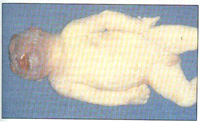
Fig. 164. Congenital anencephalia
Pachygyria — a condition in which the convolutions of the cerebral cortex are abnormally large. Thus the secondary and tertiary gyri are fully absent, sulci are short, shallow and relatively direct, the structure of cytoarchitectonics of cerebral cortex is disturbed.
Аgyria — absence of fissures, gyri, and a layer-by-layer structure of cerebral cortex in the cerebral hemispheres. Clinical signs: failure of swallowing, muscular hypotension, spasms, mental underdevelopment. The majority of children die within the first year of life.
Мicrocephalia — reduction of weight, sizes and histological structures of the brain. The primary and secondary microcephalia are distinguished. To primary one the hereditary forms of microcephalia refer, to secondary one — microcephalia developed due to organic damage of the brain of various etiology. The most frequent reasons of secondary microcephalia are the infections during pregnancy, (toxoplasmosis, rubeola, cytomegalia), various intoxications, hormonal disturbances, hypoxia.
Congenital defects of development of the olfactory analyzer — arrhinencephaly (aplasia of olfactory bulbs, furrows, tracts). More often it is encountered as a component of hereditary syndromes of multiple defects.
Congenital defects of development of medulla oblongata — aplasia and hypoplasia of pyramids and olives, which usually accompany severe disturbances of telencephalon.
The defects of cerebellum development — hypoplasia, structural changes — are seldom encountered.
The defects of development of spinal cord and spinal column — most frequent are dysraphia (spina bifida) (Fig. 165), which are connected with nonclosure of medullar tube. Usually these defects refer to back parts of spinal column, when the arches and spinal processes are absent. In the area of defect the spinal cord is usually deformed and it turns out to be open or located directly under soft tissues (muscles, skin), which it often accretes with. There are also cystic clefts of spine, when in the area of cleft a hernia sack forms, which wall is presented with skin and cranial pia mater, and contents — with a cerebro-spinal liquid. In the lumbar and sacral parts of the spinal column there can be latent clefts of vertebra. Thus hernial diverticula are absent, and the defects are closed by unchanged muscles and skin. Dysraphia and other severe damages of spinal cord are accompanied by failure of activity of anal and urethral sphincters, muscle hypoplasia of lower extremities and talipes, trophic disorders.
Аmielia — a complete absence of the spinal cord with preservation of dura mater, and spinal ganglia. Amielia is usually combined with аnencephaly.
Hydromielia — hydrocephaly of the spinal cord.
Syringomielia — the presence in the spinal cord of cavities of different size. The walls of cavities are formed by glial tissue. The cavities are usually located in the area of the neck part of the spinal cord and have a tendency to spread, thus resulting in destruction of the brain structures, and progressing gliosis round the central canal.
Diplomyelia — doubling of the spinal cord in the area of cervical or lumbar thickening. Rarer the whole spinal cord is doubled. Both brains are located in one bed consisting of pia mater and dura mater, here and there connected by glial tissue, rather well developed and having all characteristic components for brain.
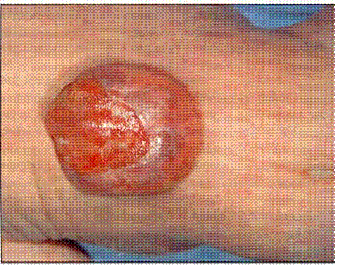
Fig. 165. Spina bifida.
Defects of Development of Cardiovascular System
Acardia — the absence of the heart is observed only at asymmetric Siamese twins.
Ectopia of the heart is its location out of the thorax. The defect is rare, usually combined with other heart defects. Cervical and extrasternal forms of defect lead to lethal outcome; at abdominal form the viability can be saved.
Dextrocardia — displacement of the heart to the right. It can be observed at an opposite location of all organs, less often as an isolated defect.
Right-side arch of aorta — the most frequent anomaly of all defects of arch. In 20-25 % of cases it is combined with Fallot’s tetrad. In isolated form it does not cause the disturbance of hemodynamics, but can manifest in dysphagia and/or respiratory impairment at squeezing of gullet or trachea.
Double arch of aorta — one arch is disposed in front of trachea, the other — behind the gullet.
Coarctation of aorta — narrowing of the isthmus, less often of pectoral and abdominal aorta. The first signs of decompensation usually manifest by 10 years of child’s life.
Aortic stenosis — pathologic narrowing of the aortic valve orifice. A semilunar valve is thus presented by a diaphragm with an orifice of various size.
Stenosis of pulmonary artery is more often valvular, rarely infundibular, usually is a component of complex heart defects.
An opened arterial defect — persisting of Botallo duct.
Aorticopulmonary fistula (aorticopulmonary defect) — fusion between aorta and the trunk of pulmonary artery as an orifice 10-30 mm by diameter, which is located above semilunar valves, and accompanied by the expressed disturbances of hemodynamics.
The atrial septal defect is one of the most frequent defects of cardiovascular system (7-25 % of all congenital defects of the heart). Primary and secondary atrial septal defects are distinguished, as well as their complete absence (cor trilocular biventriculare with one common auricle).
Lutembacher’s defect (syndrome) — a combination of congenital atrial septal defect with mitral stenosis. Usually there is hypoplasia of the left ventricle and aorta, the right half of the heart is hypertophied, pulmonary artery is extended. Decompensation may occur at any age.
Ventricular septal defect — in most cases it is a part of complex defects: Fallot’s tetrad, Eisenmenger complex, transposition of large vessels, etc.
Cor triolocular biatriatum with one common ventricle — a complete absence of interventricular septum. Cor biloculare (two-chambered heart) — absence of interatrial and interventricular septa.
Fallot’s triad — valval stenosis of pulmonary artery combined with defect of interatrial septum and hypertrophy of the right ventricle.
Fallot’s tetrad — stenosis of pulmonary artery, a high defect of interventricular septum, dextroposition of aortic ostium, " sitting" above the defect, hypetrophy of the right ventricle.
Fallot’s pentalogy — a combination of Fallot’s tetrad with defect of interventricular seprum.
Eisenmenger complex — a high defect of the membranous part of interventricular septum, aorta, which is" sitting" above the defect, and hypertrophy of the right venticle. The pulmonary artery is located and developed normally.
Common aortic trunk — safety of primary embryonic arterial trunk, as a result from the heart one vessel goes out.
Transposition of vessels — origin of aorta from the right ventricle, and pulmonary artery — from the left one. In the absence of roundabout shunts, the defect is incompatible with life.
Аtresia of tricuspid valve, Epstein’s defect, primary pulmonary hypertension, valval anomalies, other heart defects, w
|
|
|


