 |


Mode of delivery
|
|
|
|
Placenta previa is an absolute indication to the planned caesarean section.
In total placenta previa without bleeding, the expectant mother is hospitalized in the antenatal ward, where conservative treatment aimed at prolonging pregnancy is carried out, and delivery by cesarean delivery is routinely performed at the 38th week of pregnancy (before the onset of labor pains). Given the high risk of massive bleeding, the surgery should be performed in tertiary hospital using blood-saving technologies. Bleeding in total placenta previa is an absolute indication for immediate cesarean section regardless of the gestational age.
In case of partial placenta previa in parturient women with insignificant bloody discharges, it is possible to lead births through natural birth canals. With cervical dilation for 4-5 cm artificial amniotomy is indicated to stop bleeding. The rupture of the amniotic membranes facilitates the ldescending of the fetal head into the small pelvis, which presses the exfoliated portion of the placenta, stops further detachment and stops bleeding. If the bleeding continues after an amniotomy, the emergent cesarean section is performed. If childbirth was conducted through natural ways, active management of the third period is necessary.
Immediately after the birth of the fetus, manual removal of the placenta and separation of the placenta is indicated, which is mandatory and allows timely verification of the placental attachment anomaly (adherent placenta, fused placenta), uterine hypotony or rupture of the lower segment of the uterus, if any.
In the process of delivery by any method, the woman giving birth needs special attention.
Management of puerperal period after placenta previa
The puerperium in patients with placenta praevia is often complicated.
Infectious diseases may develop postpartum because the placental site is close to the vagina and the resistance of the woman is impaired after the loss of blood. The puerperant requires careful attendance and observation. Uterotonic agents and blood transfusion are prescribed if necessary. Diseases of infectious etiology should be appropriately treated.
Premature Separation of Normally Implanted Placenta
Premature separation of normally implanted placenta (syn.: Abruptio placentae, placental abruption) means its separation before the delivery of the fetus (during pregnancy, the first and second stages of labor).
It is one of the most severe complications of gestation which can lead to maternal and perinatal morbidity and death.
Incidence. It varies from 0. 05 to 0. 5%.
Etiology. The most frequent causes of placental abruption are diseases associated with vascular disorders. These are EPH-complex (preeclampsia, eclampsia), pyelonephritis, heart diseases, diabetes mellitus, and avitaminoses. Hard work, mechanical traumas, stresses, absolutely shortened umbilical cord may be of importance, especially in case of connection with vascular diseases.
Pathogenesis.
|
|
|
The pathological changes in the tissues adjacent to the uteroplacental junction are conspicuously present. The earliest lesion is degeneration and necrosis of the vessels of decidua, especially in cases associated with preeclampsia, chronic hypertension, pyelonephritis, etc. Disruption of the vessels with formation of decidual hematoma occurs. Decidual hematoma leads to placental separation. Ruptures of the basal plate also occur, thus connecting the hematoma with the intervillous space. The hematoma so formed, leads to compression and ultimate destruction of the placental unit adjacent to it. If the smaller vessels are involved and the hematoma occurs away from the margin, the entity is evident only after the expulsion of the placenta (retroplacental hematoma). The features of retroplacental hematoma are: depression found on the maternal surface of the placenta with a clot which may be found firmly attached to the area; areas of infarction with varying degree of organization.
If a major spiral artery is involved, a big hematoma is formed. As the uterus remains distended by the fetus, it fails to contract and compress the torn bleeding point. The blood so accumulated finds its way in the following direction:
Complete accumulation behind the placenta which may be separated totally up to the margin where it is adherent to the uterine wall.
Blood may dissect downwards in between the membranes and the uterine wall and ultimately escapes out through the cervix or may be kept concealed by the pressure of the fetal head on the lower uterine segment.
The blood may gain access to the amniotic cavity after rupturing through the membranes.
Thus hemorrhage in premature separation of placenta may be internal (accumulation of blood behind the separated placenta or between the membranes and the uterine wall) (Fig. 186), external (the blood comes out of the cervical canal to be visible externally) (Fig. 187), and a mixed form (Fig. 188).
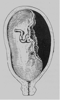
Fig. 186. Premature separation of placenta: accumulation of blood behind the separated placenta (internal hemorrhage)
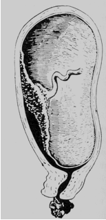
Fig. 187. Premature separation of placenta: external hemorrhage (the blood comes out of the cervical canal to be visible externally).

Fig. 188. Premature separation of placenta; mixed form of the hemorrhage.
Couvelaire uterus (utero-placental apoplexy). It is a pathological condition which is characterized by hemorrhagic saturation of the uterine wall, which may be patchy or diffuse. The uterine muscles over the affected area are necrosed and there is infiltration of blood in between the muscle bundles. (Fig. 189) Impossibility of uterine contraction, necrosis, etc., are common results of this complication.
When some mechanical factor such as trauma or sudden decompression of the uterus or short cord operates, it usually leads to marginal placental separation. The bleeding is from the torn uterine sinuses and the blood loss is proportionate to the extent of placental separation. The track downwards between the membranes and the uterine wall is to be revealed outside.
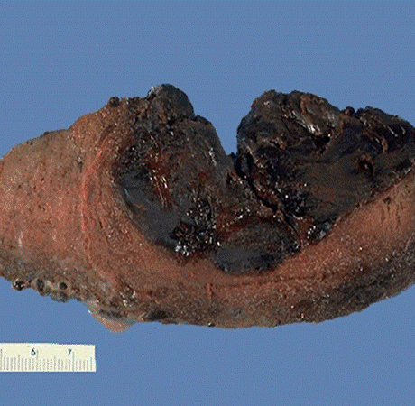
Fig. 189. Abruptio placenta with retroplacental blood clot is shown here in cross section.
There are hardly any morbid pathological changes in the uterine wall or distant organs nor there is any blood-biochemical mechanism.
|
|
|
In a severe form and if the patient is left undelivered, the pathological changes of other organs may occur, but they are more pronounced in the liver and kidneys. Blood coagulopathy is one of the commonest complications, which usually occurs in an hour after the onset of premature separation of the placenta.
Classification
There are the following options for premature placental abruption (by Radzinskiy V. E., Aleksandr М. Fuks, 2016):
• a complete placental abruption, when the detachment occurs over its entire maternal surface;
• partial placental abruption, when any part of its maternal surface exfoliates from the placental bed.
A partial detachment can be progressive and not progressive.
Depending on severity of clinical symptoms, there are:
• mild degree (detachment of a small section);
• moderate degree (detachment of 1/4 of the placenta surface);
• severe degree (detachment more than 1/3 of the placenta surface).
Clinical classification of placental abruption is based on bleeding variants:
• detachment with external bleeding (discharge of blood from the vagina);
• detachment with internal bleeding (formation of utero-placental hematoma, blood from the vagina is not allocated);
• Detachment with combined bleeding (internal and external bleeding)
Practically, these classifications are not so important because of the alterations in the organism, which depend not only on the area of separation, but on the previous condition of the maternal organism.
Total and partial separation of the placenta endangers the mother and the fetus: the woman may die from excessive blood loss and shock associated with overdistension of the uterus, while the fetus often dies from asphyxia. The danger of the fetal distress arises when a third of the placenta separates. If half of the placenta has been separated, the intrauterine fetus quickly dies.
|
|
|
