 |


Chapter 7. Ultrasonic diagnostic examination in obstretrics
|
|
|
|
Ultrasound examination (sonography/ ultrasound /echography) is a diagnostic non-invasive method of imaging internal organs of a patient by ultrasonic waves (Fig. 19, 20).
Ultrasound for diagnostic purposes in obstetric practice was first used in 1958 by Donald J. and colleagues.
Ultrasonic waves are oscillations of the particles of the material medium in the form of alternating zones of compression and extension. The frequency of ultrasonic waves is above a threshold, perceived by the human ear i. e. greater than 20, 000 Hz.
The method of ultrasound diagnosis is based on direct and inverse piezoelectric effect, open by brothers Jacques and Pierre Curie in 1880-1881.

Fig. 19. Modern ultrasound scanners.
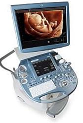

Fig. 20. Modern ultrasound scanners.
When enabled, ultrasound scanner electric current acts on the piezoelectric elements, causing the crystals to oscillate, i. e., to constrict and expand with a certain frequency and generate ultrasonic waves at the appropriate frequency.
Ultrasonic waves are characterized by:
• oscillation period - the time during which a molecule (particle) makes one complete oscillation;
• Frequency - the number of oscillations per unit of time;
• Length - the distance that 1 oscillation takes in space;
• Velocity of propagation, which depends mainly on the elasticity and density of the medium.
The wavelength is inversely proportional to its frequency. The higher the frequency, the shorter the wavelength, the higher the resolution of the ultrasonic probe (Fig. 21, 22).
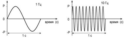
Fig. 21. The dependence of the wavelength on the frequency.
The frequency range of the ultrasonic sensors used today in medical practice for diagnostic purposes, from 2 to 20 MHz.

Fig. 22. Ultrasonic sensors (transducers).
Any medium, including body tissue, prevents the spread of ultrasound, that is, has an acoustic resistance (impedance).
The ultrasound, generated by the ultrasonic scanner, reaching the boundary between two media with different acoustic impedance is partially reflected and partially continues to spread in the new environment to the next boundary with a different impedance. Similar processes take place at every meeting of the ultrasonic wave with the medium with acoustic impedance, up to full attenuation of the ultrasonic wave (Fig. 23).
Fig. 23. Reflection and refraction of the ultrasonic waves at the interface between the media.
The sound conductivity is the ability of ultrasound to propagate to a depth. The highest sound conductivity have liquid environments. The ability of the object to reflect ultrasound termed " echogenicity. "
Echogenicity depends on:
• the magnitude of the acoustic impedance of the reflecting surface (the medium with a strong acoustic impedance will reflect more ultrasound waves, and, when applied to the piezoelectric elements of the ultrasonic sensor, generates a stronger signal, which on the screen looks brighter and lighter);
|
|
|
• the frequency of ultrasonic fluctuations (at a higher frequency most part is reflected),
• angle of permeation – the highest reflection occurs in the perpendicular direction of permeation of ultrasonic waves (this is important when performing Doppler studies).
The studied object may be hyperechogenic, may have a high, moderate, low echogenicity or be anechoic (Fig. 24, 25, 26, 27, 28).


Fig. 24. Arrow is pointing at the " ANECHOIC" bladder.


Fig. 25. Arrow is pointing on the proliferative endometrium " LOW ECHOGENICITY"


Fig. 26. Arrow is pointing on the myometrium of the uterus " MEDIUM ECHOGENICITY".


Fig. 27. Arrow is pointing on the secretory endometrium " INCREASED ECHOGENICITY"


Fig. 28. Arrows are pointing on the " HYPERECHOGENIC" bone fragments of the fetus in the uterus, which remained after the medical abortion.
Stationary and portable ultrasound scanners, operating in a variety of real-time fashion, are used in obstetrics and gynecology (Fig. 29).
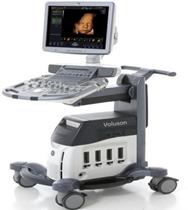
Fig. 29. The stationary ultrasound scanner

Fig. 30. Portable ultrasound scanner.
1. B-mode ( B=brightness ) - two-dimensional (2D) gray-scale image (Fig. 31) An amplitude of the reflected ultrasound signals is converted into a gray scale image.

Fig. 31. Uterine corpus.
2. D-mode (D=Doppler) - this imaging mode is based on the Doppler effect, ie. change in frequency (Doppler shift) caused by the reciprocal movement of the sound generator and the observer. The image is presented as a curve of blood flow velocity obtained by Pulsed-Wave Doppler.

Fig. 32. A curve of blood flow velocity of an artery
3. CD (Color Doppler ) – coding of moving thread a different color depending on the direction of movement to the sensor and intensity of flow (Fig. 33).

Рис. 33. Aortic arch and descending aorta of the fetus.
4. PD (Power Doppler ) is a more sensitive version of the CD, without the characteristics of the direction of flow (Fig. 34).

Рис. 34. The place of attachment of the umbilical cord of the fetus.
5. Modes of " volumetric" visualization of 3D (three-dimensional reconstruction) and 4D (3D in real time) (Fig. 35, 36).

Fig. 35. The face of the fetus.

Fig. 36. Twins.
|
|
|
