 |


Auscultation of fetal heart sounds (F.H.S.)
|
|
|
|
Table 3.
HCG levels in weeks from the last normal menstrual period (LNMP) 
The placenta produces placental laktogen — chorionic somatotrophic hormone, which has an anti-insulinic action, and due to this action, it intensifies the processes of glycogenesis in the liver, decreases tolerance of organism to glucose, and intensifies lipolysis.
The placenta also produces other hormones, such as melanocyte-stimulating hormone (MSH), adrenocorticotrophic hormone (ACTH), thyrotropic hormone (TTH), oxytocin, vasopressin, as well as biologically active substances — relaxin, acetylcholine and others.
The placenta produces the steroidal estrogenic hormones, one of which is estriol. The level of estriol in blood at pregnancy increases by 5–10 times, and excretion with urine increases by more than hundreds times. Estriol, neutralizing the action of other estrogens of placenta (estrone and estradiole), decreases the contractile activity of uterus during pregnancy.
There are great changes in the hypophysis, thyroid and adrenal gland too.
Thus, the detection of growth of luteinizing hormone (LH) of hypophysis may be one of the first signs of pregnancy. Usually the production of melanocyte-stimulating hormone (MSH) increases as well, that determines predisposition to hyperpigmentation at pregnants.
The anterior lobe of the hypophysis produces hormones which stimulate the function of the yellow body at the beginning of pregnancy. The posterior lobe of the hypophysis produces vasopressin and oxytocin. Oxytocin stimulates contraction of uterine muscles, probably, due to facilitation of prostaglandin action. Oxytocin has a weak antidiuretic action; it has an isolated vasodilative action as well, although during pregnancy estrogens suppress this action. Accumulation and action of oxytocin are found to be in direct dependence on maintenance of estrogens and serotonin in placenta, which block oxytocinase. This enzyme inactivates oxytocin in the blood of woman during pregnancy; it appears in trophoblast, sometimes it is called the enzyme of pregnancy.
The production of pituitrin in the posterior lobe of hypophysis considerably increases at term of delivery. In postpartum period, the hormones of anterior lobe of hypophysis make for becoming of a new function — function of lactation.
During the first months of pregnancy some rise of thyroid function can be marked, in the second half sometimes its hypofunction occurs. The level of circulatory thyroxin during pregnancy does not rise on the whole, although intensity of basic exchange increases by 10% as compared with initial. Clinically a slight swelling of thyroid may be found at pregnant, caused by the rise of its activity because of necessity to compensate the increased excretion of iodine by kidneys.
During pregnancy, the fascicular zone (zona fasciculate) of adrenal cortex is hypertrophied. There is an increased production of glucocorticoid hormones, which regulate a carbohydrate and albuminous metabolism. Synthesis of hydrocortisone, estrogens, progesterone and androgens increases in adrenal cortex. The exchange processes in the organism of pregnant accelerate under the influence of adrenal glands, the content of cholesterol and other lipids in blood is multiplied, pigmentation of skin increases.
|
|
|
The level of aldosterone which stimulates the excretion of sodium by kidneys rises at normal pregnancy.
The level of insulin increases, probably, because of stimulation of Langerhans’ islets by lactogenic hormone of placenta.
The Cardiovascular system
The cardiovascular system of healthy women significantly changes during normal pregnancy.
Changes of Hemodynamics
Hemodynamic changes in pregnancy are compensatory adaptation to the co-existence of maternal and fetal organisms. They are manifested by increase of cardiac output, blood volume, pulse rate and venous pressure. The changes of hemodynamics are connected with the increase of body weight, enlargement of uterus, fetus, placenta, acceleration of metabolism by 15–20%, development of additional placental blood circulation. The physiological hypervolemia is one of the main mechanisms which facilitate the optimal level of microcirculation in the placenta and vital organs of mother (heart, brain, kidneys) during pregnancy and labor. The volume of blood plasma in pregnancy begins to increase from the 10th week of pregnancy. The intensive increase of volume of circulating blood plasma proceeds to 34th week of pregnancy, after that the increase proceeds, but considerably slower. On the 34th week of pregnancy, the increase of plasma quantity reaches about 30-40%, at the end of pregnancy — 50%. Thus, the volume of circulating plasma by the end of pregnancy makes up 3, 900–4, 000 ml. The amount of red blood cells (RBC) is also multiplied, but to a lesser degree, increased by the end of pregnancy approximately by 18-20% in comparison with the initial level. The disproportional increase in plasma and RBC volume produces a state of hemodilution during pregnancy at term of 26-32 weeks of gestation. Thus, even though the total hemoglobin content and RBC count increase during pregnancy to the extent of 18-20%, there is an apparent fall in hemoglobin concentration approximately to 10-20%. As a result, oligocythemic anemia and decrease of blood viscosity occur. Therefore, the relative decrease of hemoglobin volume, called “hydremia of pregnancy”, or physiological hypervolemia (autohemodilution), usually takes place almost in every pregnant woman. Administration of ferrotherapy may prevent this condition.
The physiological hypervolemia is an important compensatory-accommodative mechanism, which helps:
• to provide for optimal level of microcirculation in vital organs and placenta during pregnancy;
• to permit some pregnant to lose about 30–35% of blood volume without development of expressed hypotension (protective action of autohemodilution).
The average arterial pressure at pregnancy rises from 95 mmHg in non-pregnant to 105 mmHg, that facilitates the oxygen transport from mother to fetus. The average blood pressure is usually determined by formula:
ABP = SBP + 2DBP
------------------------ 3,
ABP = SBP + 2DBP
where:
ABP — average blood pressure,
SBP — systolic blood pressure,
DBP — diastolic blood pressure.
|
|
|
The cardiac output, which in non-pregnant women makes up approximately 4. 2 litres per minute, rises to 6. 5 litres per minute, beginning with the 8-10th weeks of pregnancy. This level remains static until term, but just before the beginning of labor there is a tendency to decrease of cardiac output. The increase of cardiac output is mainly affected by increase of stroke volume and increase of pulse rate from 72 to 78.
The minute cardiac volume in normal pregnancy increases on average by 30-32% to 26-32 weeks of gestation. At the end of pregnancy, the minute cardiac volume slightly decreases, but to the onset of labor it increases and even exceeds the initial level.
The peripheral resistance decreases, especially in the middle of pregnancy, so there is a tendency to arterial hypotension between the 16th and 28th weeks of gestation. The decrease of common and peripheral resistance of vessels is explained by formation of utero-placental circle of blood circulation. Moreover, vasodilatory action of progesterone and estrogens may affect it.
The arterial pressure, if not to speak about a tendency to a slight fall in the middle of pregnancy, practically does not change at healthy pregnant. The individual level of blood pressure is determined by correlation of four factors:
• decreased common peripheral resistance;
• decreased viscosity of blood;
• increased volume of circulating blood;
• increased minute cardiac volume.
The first two factors contribute to the decrease of blood pressure, the last two — to its increase.
Interaction of all four factors maintains blood pressure at the optimal level.
Cardiac activity
There is a physiological tachycardia in normal pregnancy. In the 3rd trimester of pregnancy the pulse rate is increased by about 15 beats per minute. The central venous pressure is raised on average from 2-5 cm of water in non-pregnant state to 8-10 cm of water in pregnancy. The pressure in the veins of upper extremities does not change. The pressure in veins of lower extremities increases. Partially it is due to force of gravity, partially — due to obstacle to backflow of blood from uterus and placenta. The gravid uterus produces a compression effect on the inferior vena cava, when the patient is in supine position. Worsening of vein outflow through the lumbar and paravertebral veins, and reduction of minute cardiac volume cause a collapse in some women. Therefore, the position on the back is not recommended for pregnants.
The high standing of the uterus results in limitation of diaphragm mobility and change of heart position in the thorax. Therefore, in half of healthy women during pregnancy a systole murmur may be audible in the apical area of the heart. Strengthening of the first tone at the apex of the heart is marked; sometimes its accent is observed on the pulmonary artery. The significant changes of electrocardiography during pregnancy are not observed.
Hematologic Changes in Pregnancy
The content of hemoglobin due to autohemodilution in the III trimester of pregnancy goes down from 139 to 112 g/l and lower (Table 3).
The total number of leucocytes is multiplied from 7, 500 to 10, 000 per mm3, and the ESR (erythrocytes sedimentation rate) amounts to 50 mm during the first hour.
The quantity of thrombocytes is approximately doubled running by the term up to 316, 000 per mm3. The content of serous fibrinogen increases from 3 g/l in non-pregnant to 6 g/l to the moment of birth. In the II and III trimester of pregnancy the content of coagulate factors in blood increases, the prothrombin index rises. The rate of blood coagulation gradually increases; the structural properties of blood clot are enhanced.
The total number of lipids increases from 6 to 9 g/l mainly due to cholesterol.
The level of albumens diminishes from 70 to 60 g/l that causes the falling of osmotic pressure of plasma, leading to edema. The albumen/globulin coefficient falls from 1. 5 to 1. These changes take place due to decrease of albumen level and increase of content of alpha- and beta-globulins. The level of gamma-globulins goes down.
|
|
|
Table 4
Normal Hematological Indexes in Pregnancy
| Index | Non-pregnant state | I trimester of pregnancy | II trimester of pregnancy | III trimester of pregnancy |
| Hemoglobin (g/l) Hematocrit (l/l): arterial venous Erythrocytes (х1012/l) Leucocytes (х 109/l) Neutrophiles (%) Basophils (%) Eosinophiles (%) Lymphocytes (%) Monocytes (%) ESR (mm/h) | 139 (115-160) 0. 35 0. 40 4. 2 –5. 4 7. 4 0. 5 2. 0 38. 0 4. 0 | 131 (112-155) 0. 33 0. 36 4. 2 — 5. 4 10. 2 0. 2 1. 7 27. 9 3. 9 | 120 (108-144) 0. 36 0. 33 3. 5 — 4. 8 10. 5 0. 2 1. 5 25. 2 4. 0 | 112 (110-140) 0. 34 0. 32 3. 7 — 5. 0 10. 4 69. 6 0. 1 1. 5 25. 3 4. 5 |
The Respiratory system
Pregnancy requires intensification of respiratory function to meet the increasing metabolic demands of growing fetus. Therefore, beginning with the 8-9th week of pregnancy, the respiratory system of mother undergoes a number of morphologic and functional changes, which provide the supply of oxygen and excretion of carbon dioxide in accordance with needs of organism.
The morphologic and functional changes of the thorax
By the end of pregnancy, the diaphragm goes 4 cm up, and, in spite of this, its excursions in the process of breathing have a large scope both in a vertical and horizontal position.
The increased mobility of diaphragm is provided by diminished tone of abdominal musculature and expansion of the thorax, its circumference being 6 cm larger due to increase of the transverse diameter. The changes of thorax and diaphragm cause changes of the respiratory type in pregnant, which becomes mainly diaphragmatic.
Pulmonary Ventilation
During pregnancy, the activity of lungs is increased in connection with growing demands for oxygen. Total consumption of oxygen at the end of pregnancy increases by 30–40%, and during the 2nd stage of labor — by 150–250% of the initial consumption, achieving 800–900 ml O2/min in primigravida.
Hyperventilation during pregnancy causes a fall of arterial PCO2 from 38 to 32 mmHg. These changes facilitate the transfer of CO2 from the fetus to the mother.
These compensatory reactions are provided by processes of lung hyperventilation, heart hyperfunction, activation of erythropoiesis leading to increased number of RBC.
However, the excursion of diaphragm during pregnancy remains somehow limited and pulmonary ventilation — laboured. It is manifested by accelerated breathing (by 10% of initial one), and gradual increase of respiratory volume (at the end of pregnancy — by 30-40% of initial volume). The minute respiratory volume rises from 8. 4 litres per minute on the 12th week of pregnancy to 11. 1 litres per minute to the term of labor.
The increase of respiratory volume happens due to decrease of expiratory reserve volume.
The vital capacity of the lungs (maximum volume of air that is breathed out by maximum expiration after maximum inspiration) does not change during pregnancy in fact.
In spite of the fact that the vital capacity of the lungs does not undergo the significant changes during pregnancy, its components — current volume and inspiratory reserve volume — undergo the essential quantitative changes. The current volume, i. e. quantity of inhaled and exhaled air on ordinary breathing, progressively increases from the third month to the term of delivery, becoming 100–200 ml (40%) larger than in non-pregnant. The inspiratory reserve volume increases during the late terms of pregnancy due to increase of thorax size. The inspiratory reserve volume in combination with the current volume make up the capacity of inspiration, which increases on the 6th — 7th months of pregnancy by 120 ml (5%) in comparison with non-pregnant. As opposed to this, the expiratory reserve volume falls down approximately by 100 ml (15%) in the second half of pregnancy, achieving the least values on the 24–28th week. The reduction of expiratory reserve volume is explained by the rise of current volume, and as a vital capacity does not change, at the end of ordinary exhalation the compression atelektasis of the lungs in pregnant increases, therefore the lungs contain comparatively less air than lungs of non-pregnant woman.
|
|
|
Residual volume — quantity of air remained in the lungs after maximum exhalation — decreases in pregnancy by about 20%. The functional residual capacity of the lungs and total lung capacity are reduced because of high standing of diaphragm.
Maximum capacity of the lung — volume of air contained in the lungs by the end of maximum inhalation — is decreased.
The work of respiratory muscles increases, their consumption of oxygen increases, although the resistance of respiratory tract by the end of pregnancy goes down almost by 1. 5 times.
Mechanical properties of the lungs. General resistibility of lungs is 50% less in pregnancy; it is probably due to weakening of smooth muscle tone of bronchioles depending on progesterone action.
Perfusion of lungs during pregnancy increases, diffusion of oxygen through an alveolar-capillary membrane does not change, or slightly decreases, preserving the capacity to increase on loading.
Thus, the morphologic and functional changes of the respiratory system during pregnancy create the necessary conditions for realization of pulmonary hyperventilation, which, in combination with the rise of pulmonary perfusion and increase of an exchange alveolar-capillary area, makes it possible to intensify the respiratory exchange of gases according to the needs of organism of a pregnant and her growing fetus.
The Urinary system
In the first and second trimester of pregnancy the blood circulation in kidneys is intensified, gradually returning to the initial level by the term of delivery. In the late terms of pregnancy the enlarged uterus hinders in the venous outflow from kidney, although it is revealed only when the pregnant lies on the proper side.
The intensity of glomerular filtration rises by 50%, returning to normal only after delivery of fetus. The inulin clearance increases from 90 to 150 ml/min. Almost 100 liters of liquid is filtered daily. In spite of this, the excretion of urine decreases insignificantly. During the II trimester of pregnancy the increase of cardiac output, volume of plasma, speed of glomerular filtration up to 40% are observed. In the III trimester of pregnancy, these indexes return to the initial level. During the last 3 months of pregnancy the blood circulation in kidney is 10% higher as compared to normal level, while glomerular filtration returns to normal by the end of the VIII month of pregnancy.
Because of the increased glomerular filtration and increased volume of plasma, the level of creatinine in the blood serum is lower than in non-pregnant women. It is also promoted by reduction of albumen catabolism during pregnancy.
The excretion of urea and uric acid increases too.
The kidney threshold for glucose sharply decreases at term of 16–20 weeks of pregnancy, which frequently causes glucosuria. The excretion of 140 mg of glucose daily is an upper limit of physiological glucosuria.
During pregnancy approximately at 20% of women the occurrence of orthostatic proteinuria is marked. The basic indexes of kidney function are presented in Table 4.
Under the action of progesterone the muscular fibers of urinary bladder are hypertrophied, the latter becoming elongated and flaccid, that may result in stagnation of urine.
Due to relaxing effect of progesterone on smooth musles some atony of the ureters may be observed that may result in vesicoureteral and pyelorenal reflux (reflux pyelorenalis). This process is aggravated with the growth of uterus which presses the urinary bladder, thus promoting the development of infection and hydronephrosis. So, the conditions for development of pyelonephritis during pregnancy are created, the risk of its occurrence being especially great at disturbance of vaginal ecology.
Table 5
Function of Kidneys at Pregnancy
|
|
|
| Index | Non-pregnant women | I trimester of pregnancy | II trimester of pregnancy | III trimester of pregnancy |
| Blood urea nitrogen, mmol/l Creatinin, mmol/l Urates, mmol/l Renal blood flow, ml/min Clearence of creatinine, ml/min Protein, g/daily Glucose, mmol/daily Amino acids, mmol/l | 2. 5 — 6. 5 0. 279 964 ±272 90–120 0 — 0. 03 - 5. 68 | 3. 5 - 1280±161 110–150 0 — 0. 03 - 11. 6 | 3. 3 0. 190 1216±108 - 0 — 0. 04 - 15. 9 | 3. 1 0. 198 940±186 109–160 0 — 0. 05 up to 7. 7 15. 5 |
Genital organs
In the reproductive system the uterus undergoes the basic changes. To the moment of birth the uterus is enlarged in size up to 28х24х20 cm. Thus, the length of unpregnant uterus makes up 7-8 cm; by the end of pregnancy it increases up to 37-38 cm. The transverse size of the uterus increases from 4-5 cm out of pregnancy to 25-26 cm in full term pregnancy. The mass of uterus increases from 50-100 g out of pregnancy to 1, 000–1, 500 g to the moment of birth.
The increased sizes of uterus are caused rather by hypertrophy of muscular fibres than by increase of their number. Each muscular fibre lengthens by 10-12 times and thickens by 4-5 times. Hypertrophy occurs due to action of estrogens and progesterone.
The lower segment of the uterus begins to form approximately from the 12th week of pregnancy partly from the lower part of uterus body and partly from the upper part of the cervix, which is covered by a glandular epithelium similar to epithelium of body of uterus; the cervical canal slightly shortens. The cervix of uterus becomes softer and more vascularized, acquiring a cyanotic color. The cervical canal remains densely closed by mucus plug, which serves as a barrier on the way of bacteria from the vagina. The epithelium of the cervical channel overgrows; the glandular tissue becomes more active.
A muscular layer of the uterus becomes soft, plastic and more elastic. The uterus acquires the ability to respond by strengthening of the tone to different irritations. The mucous membrane of the uterus undergoes characteristic alterations; a decidual layer develops from a functional layer of endometrium.
A vascular network of uterus overgrows: the arterial, venous, lymphatic vessels increrase in quantity, become broader and longer. Blood vessels in the area of attachment of placenta overgrow significantly. The number of nervous elements of uterus is multiplied; the new sensible receptors providing the transmission of nervous impulses appear.
Excitability of uterus in the first months of pregnancy decreases. However, the normal rhythmic contractions of uterus, characteristic of a lutein stage of menstrual cycle, increase gradually, although they remain quite painless (Braxton-Hicks contractions). In the course of pregnancy these contractions gradually increase in intensity and frequency and, though they are not strong enough to cause opening of the uterus cervix, they may be of certain significance for “ripening” the cervix of uterus.
There is a progressive increase of quantity of contractile protein, called actomyosin, in the uterus muscles; the level of total phosphorus is multiplied; accumulation of creatine phosphate, glycogen occurs. Such biologically active substances as serotonin, catecholamines, histamine gradually accumulate. The ligaments of uterus elongate and thicken, that makes for holding the uterus in a correct position both during pregnancy and in labor. The round ligaments of uterus, sacrouterine ligaments undergo the greatest hypertrophy.
The fallopian tubes thicken due to serous impregnation of tissues. In the course of pregnancy the uterine tubes descend along the lateral surfaces of uterus, becoming inactive during pregnancy.
The ovaries in the course of degeneration of yellow body become inactive, the cyclic changes in them halt, with the development of pregnancy they move from the cavity of small pelvis to the abdominal cavity.
The vagina and the pelvic floor become softer; their vessels multiply in quantity. The thickness of vaginal epithelium increases as well; the reaction of vaginal medium becomes sourer.
Blood supply of the external genital organs increases; varicosity can occur on the labia majora. Elasticity and flexibility of vagina walls, external genital organs and pelvic floor increase, as a result they become more tensile, preparing for passing fetus during delivery.
Breast changes
The growth of breasts begins at puberty under stimulation of sex hormones.
During pregnancy the breasts are prepared for secretion of milk. The lobules increase in size, and fine particles of fat develop in the alveolar epithelium. The blood supply to the breasts is intensified; subcutaneous veins are dilated and shown through the delicate skin. The breasts become engorged; tingling is often felt by pregnant. The nipples increase in size, and areolar area is characterized by a greater degree of pigmentation. The smooth of the nipples become more excitable. When stimulated mechanically, the nipple muscles contract, the areolar area decreases in size and nipple becomes more projected.
Small amount of fluid (colostrum) may discharge from nipple during the early months of pregnancy.
The breasts get prepared for lactation under the action of placental hormones and lactogenic hormone of the anterior pituitary.
Gastrointestinal system
In the course of pregnancy there is some displacement of organs of digestion from the anatomical point of view. Thus, the stomach is located more horizontally, the increased pressure on diaphragm can result in disorder of activity of stomach sphincters, because of which the sour content of the stomach is belched and cause heartburn. The small intestine is displaced upwards and to the peritoneum wall. The cecum with appendage is moved upwards and sideways — a trap for a careless surgeon.
Contractility of smooth muscles of intestine diminishes, probably, under the influence of progesterone that often results in constipations. A tendency to constipations can be aggravated by the increased absorption of water in the colon. Frequently cholestasis occurs that results in a cholestatic jaundice. Acidity of gastric juice decreases.
Pregnancy at its normal course usually does not cause any significant changes in the liver. Histologically the increase of glycogen content and fatty deposits in the liver cells are revealed. The increase of alkaline phosphatase (from 26 to 75 ME against 25 ME in non-pregnant women), direct bilirubin (up to 0. 5 — 3. 0 mmol/l) is characteristic of pregnancy.
The Osteomuscular system
A relaxing action of progesterone during pregnancy also affects ligaments and joints; it is especially marked on pelvic joint that facilitates passing of the fetus through the birth canal. It is partially due to this phenomenon that flattening and extension of feet in pregnant take place. The tone of skeletal muscles slightly decreases, that can result in descent of shoulder girdle and compression of brachial plexus, causing a typical elbow paresthesia. However, the similar phenomena infrequently occur. A more frequent manifestation at pregnancy is the development of lumbar lordosis because of the necessity to counterbalance the weight of enlarged uterus. Such lordosis is aggravated if a woman wears high-heeled shoes.
The skin
The pigmentation of skin increases in pregnant; it is especially marked on the face, round the nipples and white line of the abdomen, being particularly expressed in brunettes (chloasma uterinum). This phenomenon is conditioned by the increase of quantity of circulatory melanostimulating hormone. On the abdomen and thighs the longitudinal strips 5-6 cm in length, and about 0. 5 cm in width appear. At the beginning they are rosy, but later they become pale and more dense. They are supposed to be caused by detachment of elastic layer of skin from other layers due to rise of circulatory hormone level of adrenal glands. They are called striae gravidarum. Sometimes birthmarks appear.
The work of sebaceous and sudoriferous glands is intensified.
The Nervous system
From the moment of pregnancy occurrence a flow of impulses begins to move to the central nervous system of mother causing the development in the central nervous system of the local focus of increased excitability — a gestation dominant. Excitability of the cortex before the 3–4th month of pregnancy is lowered, and then it gradually rises. Excitability of the lower parts of the central nervous system and reflex apparatus of uterus is lowered, that provides weakening of uterus and normal course of pregnancy. Before labor the excitability of the spinal cord and nervous elements of uterus increases, creating favorable terms for the beginning of labor pains. The tone of the vegetative nervous system changes; in this connection the somnolence, tearfulness, increased irritability, sometimes dizziness and other disorders are frequently observed in pregnant. Usually these phenomena gradually disappear with growth of pregnancy.
Metabolism
The basal metabolic processes and consumption of oxygen increase during pregnancy. The intensity of basal metabolism in a healthy non-expectant mother makes up approximately 2, 300 calories per day. At pregnancy the intensity of basal metabolism increases approximately by 10%, that is conditioned by the increased consumption of oxygen and activity of the fetus, thus the total energy consumption makes up approximately 2, 500 calories per day. Sum total of additional energy consumption for the whole pregnancy makes up approximately 68, 000 calories, half of which is covered at the expense of fats and one third — at the expense of carbohydrates. Proteins give only 6. 5% of energy, as they are basically used for formation of tissues.
The protein substances, necessary for satisfying the requirements of growing fetus in amino acids, accumulate in the woman’s organism.
Changes of carbohydrate metabolism cause the accumulation of glycogen in cells of liver, muscles, uterus, and placenta. Carbohydrates pass to fetus in a form of glucose providing the energy supply of fetus, processes of anaerobic glycolysis.
Concentration of neutral fat, cholesterol and lipids rises in blood of pregnant. Fats pass to fetus in a form of glycerin and fatty acids, which are used as energy material, and for formation of tissues.
Normally the total increase of body mass during pregnancy makes up 12 kg. One third of this, i. e. 4 kg is gained in the first half of pregnancy, and the rest two thirds, i. e. 8 kg — in the second half. 60% of total increase of body mass is caused by retention of water due to accumulation of sodium. The retained water is distributed in the following way: 1. 3 l — in plasma, 2. 0 l — in fetus, placenta and amniotic fluid, 0. 7 l — in uterus, mammary glands, and 2. 5 l — in extragenital interstitial fluid. To the moment of labor the fetus and amniotic fluid in common weigh about 5. 5 kg, and this mass is lost after birth. The remaining 6. 5 kg fall to the share of uterus, mammary glands, fatty deposits (especially in thighs and buttocks).
Thus, all changes occurring in the female body during pregnancy are physiological; they facilitate the normal growth of fetus without causing any harm to the expectant mother. Moreover, pregnancy gives a new impetus to the bloom of the woman.
Self Test
1. Which of the following blood indexes are abnormal for pregnancy?
A. haemoglobin — 124 g/l
B. fibrinogen — 4. 9 g/l
C. total albumen — 62 g/l
D. thrombocytes — 8х106
2. The level of chorionic gonadotropin by the 7th week rises to:
A. 80, 000–100, 000 mIU/ml
B. 2, 500–5, 000 mIU/ml
C. 10, 000-20, 000 mIU/ml
D. 120, 000-150, 000 mIU/ml
3. Hypervolemia in pregnancy is:
A. a symptom of disturbed blood circulation in pregnancy
B. an important compensatory-adaptive mechanism
4. The cardiac output in pregnancy is
A. increased.
B. decreased.
C. permanent.
5. Maximum capacity of the lungs in pregnancy is
A. increased.
B. decreased.
C. permanent.
6. Glomerular filtration in pregnancy is
A. increased.
B. decreased.
C. permanent.
7. To the moment of birth the uterus mass is increased to:
A. 300-500 g
B. 800-900 g
C. 1, 000 — 1, 500 g
D. 2, 000-3, 000g
8. The basal metabolism and consumption of oxygen during pregnancy is:
A. increased
B. decreased
C. permanent
9. Excitability of uterus in the first months of pregnancy is:
A. increased
B. decreased
C. permanent
10. The level of hemoglobin during pregnancy is:
A. increased
B. decreased
C. permanent
CHAPTER 10. Obstetric EXAMINATION
I. Interrogation of the patient
It consists of identification of the patient, determination of the chief complaints, social history, family history (hereditary history), past illnesses, menstrual function, sexual (genital) function, obstetrical anamnesis.
1. Identification of the patient: name, surname, age, home address, and marital status.
2. Chief complaints: what is bothering the patient, the description of the onset of the present illness and complaints at the time of examination.
3. Social history: her accommodation, living conditions, occupation, etc.
4. Family history: all data about mother, father, sisters of the patient, their illnesses, especially, hereditary illnesses (diabetes, hypertension, tuberculosis, nervous diseases, mental diseases, cancer, and familial tendency).
It is necessary to find out everything about her harmful habits: use of alcohol, tobacco, sedatives or other medications.
5. Past illnesses: children diseases (measles, epidemic parotitis, and tonsillitis). Extra-genital diseases: tuberculosis, anemia, heart diseases, diabetes mellitus, nephritis and others.
It is necessary to know all cases of injuries, operations, hospitalization of the patient during her life.
6. Menstrual function: date of onset, interval, regularity, duration, amount of flow, date of last period. Complications, if any: dysmenorrhea, menorrhagia, irregular bleeding.
7. Sexual function: the beginning, regularity, and contraceptives.
8. Obstetrical anamnesis: the number of previous pregnancies, abortions, miscarriages — their results and complications, if any.
The number of children, their age, health. The date of last labor, the course, complications, weight of the newborn baby.
II. General physical examination if the patient
1. General examination: weight, height, temperature, pulse, respiration, and blood pressure.
2. General inspection: posture, frame, constitution, and nutritional state. Emotional state in response to illness.
3. Skin complexion: turgor, pigmentation, eruption, and lesions.
4. Lymph nodes: anterior and posterior cervical, supraclavicular, and others.
5. Thyroid: enlarged, nodular, bruit.
6. Chest and lungs: character and intensity of breath, sounds, duration of inspiration and expiration, rate and rhythm of expansion.
7. Cardiovascular system: pulse rate, rhythm, volume, state of vessel wall. The measurement of maternal blood pressure is of great importance in pregnancy. It is expedient to measure it in the supine position as pressure from a gravid uterus on the inferior vena cava impedes venous return resulting in a falsely low blood pressure. This is often referred to as the supine hypotensive syndrome. The correct position is “semi recumbent” — a 45˚ tilt. The maternal arterial blood pressure should be obligatory measured in both hands, especially in the 2nd half of pregnancy.
Percussion: borders of the heart.
8. Abdomen: inspection (contour, striae graviadarum, dilated veins). Undue enlargement of the uterus, condition of the abdomen skin for availability of ringworm or scabies and any scar marks on the abdomen.
9. Uterus: to determine whether the uterine ovoid is longitudinal, transverse or oblique; whether the uterus fundus is convex or concave; whether the uterus has a flattened anterior wall or whether it is cylindrical or spherical in shape.
III. Special obstetric examination
It consists of external obstetrical examination, internal (vaginal) examination and additional methods of investigation (hematological, immunological, histological, cytological, ultrasound and others). Blood test, urine test, Wassermann test, examination of blood group and Rh-factor are necessary for all pregnants. Vaginal discharge should be analysed too (microscopy and bacteriology).
The external obstetrical examination
External pelvimetry
By external pelvimetry one should measure:
• Interspinous diameter, i. e. distance between two anterosuperior iliac spines (normally — 25-26cm);
• intercristal diameter, i. e. distance between the most widely saparated points on the outer border of the iliac crests (28-29cm);
• intertrochanteric diamater, i. e. distance between the trochanters on both sides (31-32cm);
• external conjugate, i. e. distance between the 5th lumber spine and pubic symphysis (20-21cm). (Fig. 56).

Fig. 56. External pelvimetry.
The external pelvimetry helps to imagine the internal volume of the true pelvis.
Solovyov’s index
It is a circumference of radiocarpal joint, normally equal to 14-15 cm; it can help to imagine the internal volume of the true pelvis: if this index is less than 14 cm the bones are thin — the thinner the bones, the bigger the internal volume.
Palpation of the abdomen
It can help to estimate the condition of the abdominal wall, tonus of the uterus, lie, presentation, position and type of the position of the fetus.
Lie of the fetus
Lie of the fetus is a relation of the long axis of the fetus to the long axis of the mother. It may be longitudinal, transverse, and oblique. The commonest lie is longitudinal (99. 5%).
If a long axis of the fetus corresponds to the mother’s long axis, such lie is called longitudinal and it is a normal one. If a long axis of the fetus makes up a right angle with the maternal spine, such lie is called a transverse one.
If a long axis of the fetus makes up an acute angle with the material spine this lie is called oblique. The transverse and oblique lies are abnormal and may lead to complications of labor.
Presentation
Fetal presentation is determined by the portion of the fetus which occupies the lower pole of the uterus and can be felt through the abdomen and through the cervix. Presentation may be cephalic (96. 5%), breech (3-4%), or shoulder and others (0. 5%). When more than one part of the fetus presents, (for example, head and hand, head and umbilical cord, buttocks and cord) we speak about the compound presentation. The compound presentations are pathological, usually they are connected with the transverse and oblique lie of the fetus.
Positions of the fetus and their types
Position of the fetus is the relation of the fetus’ back to mother’s left or right side. Thus, the position may be left (syn.: first) — the fetus’ back is to the left side of mother’s body (Fig. 57). The position may be right (or second) — the fetus back is to the right mother’s side (Fig. 58).
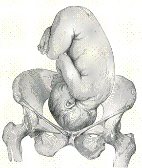
Fig. 57. The first (left) position of the fetus (the fetus’ back is to the left side of mother’s body)

Fig. 58. The second (right) position of the fetus (the fetus back is to the right mother’s side).
Type of the position is the relation of the fetus’ back to the maternal anterior abdominal wall, and is either anterior (fetus’ back is to mother’s anterior abdominal wall) or posterior (fetus’ back is to mother’s spine).
There are four special obstetrical grips which are used for external palpation (methods of Leopold-Levitsky).
The 1st grip (syn.: fundal grip) is palpation of the uterus fundus to understand the fetus’ part which is situated here (Fig. 59). If it is the head, it can be felt as a round massive ballotable part. If it is the breech, one can feel it as a massive, rather soft, non-ballotable part, without clear contours.
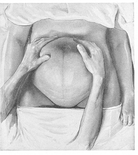
Fig. 59 The first obstetric grip (Leopold’s maneuver)
Thus, this grip helps to ascertain:
• the fetal lie — a massive part of the fetus in the fundus of the uterus is suggestive of longitudinal lie;
• the fetal presentation — breech in the fundus is suggestive of the head presentation, and vice versa.
This grip can help to understand the size of the uterus, whether it corresponds to the term of pregnancy or not, its tonus, irritability, tenderness. The palpation is done facing the patient’s face. The whole of the fundal area is palpated using both hands laid flat on it to find out which pole of the fetus is lying in the fundus: broad, soft and irreqular mass suggestive of breech, or smooth, hard and globular ballotable mass suggestive of the head.
In transverse lie, neither of the fetal poles is palpable in the fundal area.
The 2nd grip (syn.: lateral or umbilical grip) is the palpation done facing the patient’s face. The hands are to be placed flat on either side of umbilicus to palpate, one after another, the sides and front of the uterus to find out the position of the back, limbs and the anterior shoulder. The back is suggested by smooth, convex and resistant feeling. The limb side is comparatively empty and there are small knobs.
The 2nd grip can help to appreciate the lie, position and type of position of the fetus. It can help to ascertain fetal movements, uterine tone, condition of the round ligaments (Fig. 60).

Fig. 60. The second obstetric grip (Leopold’s maneuver 2)
The 3rd grip is used to appreciate the presentation of the fetus.
The examination is done facing the patient’s face. Four fingers of the right hand are placed over the lower pole of the uterus keeping the ulnar border of the palm on the upper border of the symphysis pubis; thumb is placed to the right from the middle line and other fingers — to the left. When the fingers and the thumb are approximated, the presenting part is grasped distinctly, if not engaged, and also the mobility from side is tested. (Fig 61)
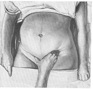
Fig. 61. The third obstetric grip: palpation of the presenting part of the fetus.
The 4th grip is pelvic (Fig. 62). Usually this grip is added to the 3rd grip. The examination is done facing the patient’s feet. Four fingers of both hands are placed on either side of the middle in the lower part of the uterus and parallel to the inguinal ligament. The fingers are pressed downwards and backwards to palpate the part occupying the lower pole of the uterus.
This grip helps to ascertain the engagement of presenting part, whether there is convergence of fingertips or not. If the fingertips convergence is not possible — the presenting part is engaged into the pelvic cavity. If the fingertip convergence is possible — the presenting part is not engaged yet.
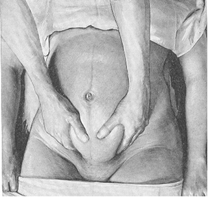
Fig. 62 The forth obstetric grip: helps to ascertain the engagement of presenting part

Fig. 63. Examples of fetal occiput presentations in relation to front, back, or side of maternal pelvis, wi9th internationally used abbreviations. The lie is longitudinal, presentation is cephalic (occipital).
Auscultation of fetal heart sounds (F. H. S. )
Auscultation of the fetal heart is one of the routine methods of assessment of fetal well-being during pregnancy. The stethoscope, invented by Laennec for chest auscultation in 1821, was used by Kergaradec to auscultate the fetal heart in 1819. Fetal heart tones are heard with an obstetrical stethoscope beginning of the second half of pregnancy (18-20 weeks with less), and with each month becomes clearer. Obstetrical stethoscope (aka Pinard horn, or Pinard stethoscope, Fig. 64) is a tool used to listen the heart rate of a fetus during pregnancy. The horn is often made of wood or metal and is hollow. It is about 20, 32 cm. long. It functions similarly to an ear trumpet by amplifying sound. By placing the larger opening firmly on the mother’s abdomen, the user can listen for the baby’s heartbeat from the smaller end.

Fig. 64. Pinard stethoscope
|
|
|
