 |


Speculum examination. The vaginal examination of the pregnant. Bimanual examination. Ultrasonic examination
|
|
|
|
Speculum examination
There are several types of vaginal specula, but more often bivalve (Cusco’) (Fig. 67) and spoon-shaped (Sims’) specula are used (Fig. 68). Both may be used during pregnancy, but in postpartum period spoon-shaped specula are more useful.
Speculum examination permits visualization of the vagina and the cervix (Fig. 69). One should pay attention to the colour and condition of the vaginal mucosa. The blue-red passive hyperemia is characteristic of pregnancy. A dilated cervix may sign the threatened abortion or premature labor. Evaluation of cervical and vaginal lesions can be accomplished by performing a Pap smear (Papanicolaou’s test), microscopic and bacteriologoical examination of any discharges.

Fig. 67 Cusco’ speculum

Fig. 68. Sims’ speculum
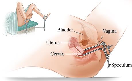
Fig. 69. Pelvic exam with speculum
The vaginal examination of the pregnant
The examiner introduces the middle and the index finger of the right hand into the vagina so that the thumb is raised upwards while the fourth and the fifth fingers are bent and rest against the perineum. The fingers palpate the muscles of the floor, the vaginal wall, the vaginal fornices, the cervix (the shape, consistency, direction), and the external os of the cervical canal (if it is opened or closed, oval or slit-shaped).
Bimanual examination
It is the basic method of obstetrical examination. The index and third finger of the right hand are introduced into the vagina, placed in the fornix, and the cervix of the uterus is then pulled slightly posteriorly. The fingers of the left hand press carefully on the abdominal wall in the direction of the small pelvis to meet the fingers of the right hand. As the fingers of both hands approach each other, the examiner palpates the uterus and outlines its position, configuration, size and consistency (Fig. 70). The bimanual examination is completed by palpating the inner surface of the pelvic bones and measuring the diagonal conjugate. This method helps to ascertain the size, consistency, shape, tone of the uterus (whether it is pregnant or not), cervical condition (opening, length, consistency, direction). The diagnosis of presentation and position of the fetus, engagement of presenting part is accurately made by internal examination. It can help to ascertain the configuration and capacity of the bony pelvis.
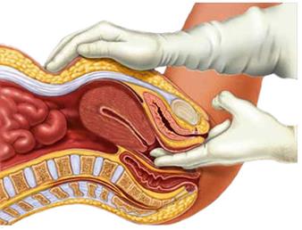
Fig. 70. Bimanual examination
IV. Additional methods of examination
Ultrasonic examination
The diagnosis of lie, presentation and position of the fetus may be difficult in the presence of marked obesity, irritable uterus, excessive liquor amnii and deeply engaged head, especially in primigravidae. Ultrasonic examination usually helps to make a correct diagnosis. It can help to ascertain the localization of placenta, its condition, size, congenital abnormalities of the fetus, to determine fetal position and size, or the reason of uterus being too large or too small for the given date of gestation. Ultrasonography is also used to detect a multiple pregnancy, a hydatidiform mole, polyhydramnios, etc. A biophysical profile (see lower) has been devised for a fetus suspected of being in distress. It is a composite of tests designed to identify a compromised fetus during pregnancy. It includes measurement of amniotic fluid volume, fetal muscle tone, fetal movement, fetal breathing, nonstress test. During ultrasound screening, the physician evaluates not only ultrasound anatomy of the fetus and excludes or detects ultrasound markers of chromosomal pathology, but also performs fetometry to clarify the term of pregnancy, assesses chorion / placental, point of attachment of the umbilical cord to the placenta, specifies the number of fetuses and determines the type of chorionicity in multiple gestation, and others. (see chapter 7).
|
|
|
Real-time ultrasonography allows direct observation of fetal and heart movements. The monitoring of the fetal heart and monitoring of fetal respiratory movement have been advocated to identify a high-risk pregnancy. Thus, biophysical profile helps to diagnose intrauterine fetal condition. Usually ultrasonography is used thrice in pregnancy: in 9-11, 16-21, and 32-36 weeks of pregnancy.
In postpartum period ultrasonic examination helps to ascertain uterine involution, retention of parts of the placenta in the uterine cavity, endometritis.
Indications for selective (if indicated) ultrasound in the first trimester of pregnancy (11-13 wk):
1. The need to confirm the existence of intrauterine pregnancy (in case of irregular menstrual cycle, using methods of assisted reproductive technology, in the period of lactation amenorrhea, etc. );
2. The discrepancy between the estimated term of pregnancy and size of the uterus (suspected ectopic pregnancy, molar pregnancy, missed abortion, to exclude anomalies of the uterus, leiomyomas, etc. );
3. Appearance of bleeding from the genital tract (to eliminate imminent abortion, abortion in progress, incomplete abortion, polyps, and others. );
4. The presence of tumor-like formations in the pelvic cavity during pregnancy;
5. Ultrasound support of medical abortion, vacuum aspiration of the ovum, chorionic villus sampling.
At the 2nd sonographic screening (at 18-21 weeks of pregnancy) the US exam is
focuses on the anatomical features of the fetus to exclude congenital malformations, the presence of a later manifestation of chromosomal pathology, assess the growth of the fetus, evaluate the amount of amniotic fluid, localization and attachment of the placenta; and assess the length of the cervix as prognostication of preterm labor. The condition of myometrium and adnexa is also controlled.
Indications for selective (if indicated) ultrasound in the second trimester of pregnancy:
1. The discrepancy between the estimated gestational term and size of the uterus (suspected intrauterine growth restriction (IUGR), undeveloping pregnancy, to avoid oligohydramnios, polyhydramnios, multiple pregnancy, etc. );
2. Ultrasonic support of multiple pregnancy, immune incompatibility, invasive techniques of prenatal diagnosis (amniocentesis, placentocentesis, cordocentesis), intrauterine medicinal- diagnostic manipulations (intrauterine blood transfusion to the fetus, in utero puncture of the fetus organs and cavities) and intrauterine operations on fetus;
|
|
|
3. The presence of vaginal bleeding;
4. The presence of tumor-like formations in the pelvic area.
|
|
|
