 |


The technique
|
|
|
|
The pregnant occupies position, lying on the back with extended feet.
One end of the stethoscope (the wide bell) is pressed on the pregnant abdomen rather tightly; the second end of stethoscope a doctor puts to the area of ear. Systematically, moving stethoscope around the belly, listen to the sounds coming from the body of the mother and fetus.
Sounds coming from the mother's body:
a) intestinal peristalsis;
b) aortic tones are synchronized with the pulse of the pregnant woman;
c) uterine noise blowing from the nature and synchronous with the pulse of the pregnant woman.
Sounds coming from the body of the fetus:
a) the fetal heartbeat, double and rhythmic, heard clearly, with a frequency of 110-170 beats per minute, do not coincide with the pulse of the pregnant woman;
b) noise umbilical cord and placental blood vessels;
c) movements of the fetal limbs, “fetal kicks”
The fetal heart sounds (FHS) are best audible through the back left scapular region, when the convex portion of the back is in contact with the uterine wall, so, it is best audible closer to the fetal head.
For optimal hearing later in the pregnancy, abdominal palpation (Leopold maneuvers) should be used to determine the position of the fetus (fetal lie, position and presentation).
Thus, to listen to heart tones of the fetus is first necessary to determine the fetal lie, position and presentation view of the position of the fetus in the uterus. According fetal lie, presentation and position the fetal heart rate is clear listened in next points (Fig. 65):
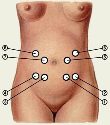
Fig. 65. Places of the most audible fetal heart tones, depending on the orientation of the fetus in the uterus: 1 - Longitudinal lie, cephalic presentation, left anterior position (left occiput-anterior presentation - LOAP), 2- Longitudinal lie, cephalic presentation, left posterior position (right occiput posterior presentation – ROPP); 3- Longitudinal lie, cephalic presentation, right anterior position (ROAP); 4 - Longitudinal lie, cephalic presentation, right posterior (ROPP); 5 - Longitudinal lie, breech presentation, left sacro - anterior (LSAP);
6- Longitudinal lie, breech presentation, left sacro – posterior (LSPP); 7- Longitudinal lie, breech presentation, right sacro – anterior (RSAP); 8 - Longitudinal lie, breech presentation, right sacro – posterior (RSPP).
In face presentation of the fetus a fetal heartbeat are audible more clearly from the side of his chest. This is due to the fact that in face presentation the head is maximally extended and the breast is adjacent to the uterine wall is closer than the back. In case of the transverse lie of the fetus, the place of the most audible fetal heart is at the level of the navel, to the right or left, depending on the location of the fetal head. In multiple pregnancy fetal heart rate usually is listened clearly in different parts of the uterus, with different rhythms and the rates (Fig. 66).
|
|
|
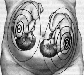
Fig. 66. Different rhythms and rates of FHS are listened in different parts of the uterus in multifetal gestation.
During childbirth when the fetal head descend in to the pelvic cavity fetal heartbeat better audible closer to the symphysis in the midline of the abdomen. Normal range of FHR is - 110-160 per minute. Rates outside of this reference range can indicate fetal distress. Normal range of fetal heart rate (FHR) is 110-160 per minute. The fetal heart rate outside this range may indicate fetal distress. Normal FHS are clear and rhythmic, do not correspond to the pulse of pregnant women. Fetal heart tones can be identified using obstetric stethoscope, ranging from 20-22 weeks of pregnancy. Auscultation of heart sounds of the fetus with a stethoscope is very subjective and depends on the skill of the doctor, and the doctor's hearing.
There are various modern methods of more accurate assessment of heart tones: cardiotocography, biophysical profile, Doppler invesigation.
Internal obstetric examination
Internal examination means speculum examination, vaginal and bimanual examination.
|
|
|
