 |


Laboratory and imaging tests
|
|
|
|
Laboratory and imaging tests are important tools helping doctors diagnose and treat health conditions. Healthcare providers use the test results along with data from physical examination and medical history to assess health status, help guide treatment decisions, track health during and after the treatment. These tests may be done in the doctor’s office, at an outpatient clinic, at a lab or in the hospital.
 Laboratory tests include blood tests, clinical urine tests, fecal tests, bone marrow test and others.A blood test is a laboratory analysis performed on a blood sample extracted from a blood vessel using a hypodermic needle, or via finger prick. Blood tests are often used to determine physiological and biochemical states, such as disease, mineral content, and organ function. Multiple tests for specific blood components are often grouped together into one test panel called a blood panel. Typical clinical blood panels include a chemistry panel or a complete blood count (full blood count, UK).
Complete blood count (CBC) is a common blood test that gives a general picture of the health. A CBC measures the number of red and white cells, platelets, and levels of hemoglobin and hematocrit in the blood. A CBC can also reflect signs of a current or recent infection, bleeding, or clotting issues. A decrease below normal in the number of red cells is called anemia. Leukocytosis (high white blood cell count) may indicate a bacterial infection. Leukopenia (very low white blood cell count) may indicate a person is at high risk for infection. Having thrombocytopenia (very low platelet count), a person may bleed or bruise more easily. The term ‘cytopenia’ or ‘pancytopenia’ means a reduction in the number of all blood cell types. Most blood counts include a CBC and leukocyte differential count (LDC) that gives the percentage of each WBC type: neutrophils, eosinophils, basophils, monocytes, and lymphocytes.
Various red blood cell indices are often reported in addition to cell counts. Automated hematology analyzer calculates the average amount (MHC) and concentration (MCHC) of hemoglobin within each red blood cell. Average RBC size (MCV) and shape (RDW) are also calculated. Automated analyzer measures MCV directly, and uses it and the RBC to calculate the hematocrit.
Blood Chemistry (complete blood chemistry) provides information about a patient’s general health. Depending on the type of panel, this test may measure the body’s electrolyte balance (sodium or potassium), protein, blood glucose, cholesterol or chemicals indicating liver and kidney function, antibodies, hormones, minerals (iron, calcium or potassium), vitamins. Doctor may also order a coagulation panel to look at how quickly blood clots or liver enzymes to determine how well the liver works, or other blood tests.
Clinical urine tests are various tests of urine for diagnostic purposes. The most common is a urinalysis, other tests are urine culture (a microbiological culture of urine) and urine electrolyte levels. A urine test is ordered to measure levels of proteins, blood cells and chemicals that may be found in the urine or to help diagnose kidney and bladder infections.
|
|
|
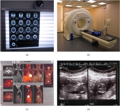
Imaging tests (radiological tests) are used to visualize the inside of the human body. Different technologies are applied to get a better picture of what’s going on in the bones, soft tissues, and organs. Imaging tests pass different forms of energy (x-rays, sound waves, radioactive particles or magnetic fields) through the body to create pictures of the scanned area. The doctor may order an imaging test "with contrast" which makes it easier to see certain organs and tissues in the body.
X-ray is a single image of the body part taken with the usage of a small amount of radiation. X-rays can depict a 2-D image of a body region, and only from a single angle. X-rays are best used to visualize hard body structures, e.g. teeth or bones.A chest X-ray widely used by doctors provides images of the lungs, heart, large arteries, ribs and diaphragm. This type of X-ray helps look for signs of disease, including infection or tumors.
CT scan (computed axial tomography) is a non-invasive test producing a 3-D image of the scanned area. It uses ionizing radiation in conjunction with a computer to create a series of images of both soft and hard tissues showing a cross-section of the body. CT creates pictures taken from different angles and produces much clearer ones than X-rays does. A CT scan may reveal signs of a tumor or injuries in the body. It is often used for soft tissue scanning, such as of the brain, the thoracic and abdominal viscera. The main disadvantage of CT scanning is patient’s being exposed to a radiation dose many times higher than that of X-rays.
MRI (magnetic resonance imaging), another type of non-invasive test used to create a 3-D image of the scanned area, uses powerful magnets and radio waves to make pictures of the body structures. MRI provides more detailed pictures than CT. It can be used to diagnose stroke and other problems involving the brain, and spinal cord, or to reveal tumors. MRI also does not expose patients to radiation.
PET scan (positron emission tomography) is a non-invasive test using a radionuclide tracer (substances emitting radiation) to provide images of the body’s inside. The main advantage is that PET can illustrate physiologic activity, including nutrient metabolism and blood flow, of the organ or organs being targeted, whereas CT and MRI can only show static images. PET is widely used to diagnose heart diseases, the spread of cancer, certain forms of infection, and other health problems.
Ultrasound (u ltrasonography) is a test using the transmission of high-frequency sound waves into the body to generate an echo signal converted by a computer into real-time images of internal organs, tissue and blood flow in the body. Ultrasound does not use radiation, so it is the least invasive of all imaging techniques. Ultrasonography is used to study heart function, certain conditions such as gallbladder disease, and fetal growth and development. The main disadvantages of ultrasonography are that the image quality is heavily operator-dependent and it is unable to penetrate bone and gas.
Also to clarify the diagnosis a doctor may order echocardiogram, electrocardiogram, mammogram, endoscopy, or other type of imaging examination.
|
|
|
Each of the tests is a piece of the puzzle and these pieces have to fit together nicely to make a complete picture of the patient’s diagnosis.
9. Correct the statements according to the text:
A. Healthcare providers use the test results to assess medical history.
B. A blood test is a laboratory analysis performed on a blood sample extracted from a soft tissue or via finger prick.
C. An increase below normal in the number of red cells is called anemia.
D. Leukocyte differential count gives the percentage of each RBC type.
E. A blood test is ordered to measure levels of proteins, blood cells and chemicals or to help diagnose kidney and bladder infections.
F. The laboratory test "with contrast" makes it easier to see certain organs and tissues in the body.
G. CT scan is a non-invasive test producing a 2-D image of the scanned area showing a cross-section of the body.
H. MRI uses powerful magnets and sound waves to make pictures of the body structures.
I. PET scan uses a radionuclide tracer to provide images of the body’s outside.
J. Ultrasound uses radiation.
10. Fill in the missing vowels:
Lab_r_t_ry, im_g_, bl__d, ur_n_l_sis, h_mat_cr_t, an_m_a, c_top_n_a, an_l_z_r, x-r_ys, _ngle, t_m_gr_phy, r_di_ti_n, ultr_s_n_gr_phy
|
|
|
